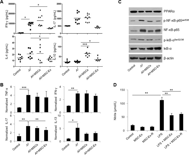
Figure 5.
MSC-Ex downregulates LN-derived Th1, Th2 and Th17 cell activation. (A) IFN-γ, IL-17, IL-4 and IL-10 levels in anti-CD3/CD28 stimulated lymphocytes from the LNs of Af-treated mice were measured by ELISA. Data are represented as the means ± s.d. (n ≥ 6 mice per group). (B) The relative expression levels of the inflammatory-associated cytokines, TNF-α, IFN-γ, IL-17 and IL-13, were evaluated by qRT-PCR in the skin tissues of AD mice. (C) The protein levels of PPARα, p-NF-κB-p65Ser536, NF-κB-65, p-IκBSer32/36 and IκB, were analyzed by immunoblotting in the skin tissue of AD mice. (D) The levels of nitrite were measured using the culture media from the basolateral side of MSC-Ex and RAW 264.7 co-cultures after 4 h of LPS (1 μg/ml) treatment and subsequent MSC-Ex (30 μg/ml) administration for 24 h. Columns represent the means of three independent experiments. Bars indicate standard deviations. *P < 0.05, **P < 0.005, ***P < 0.001.

