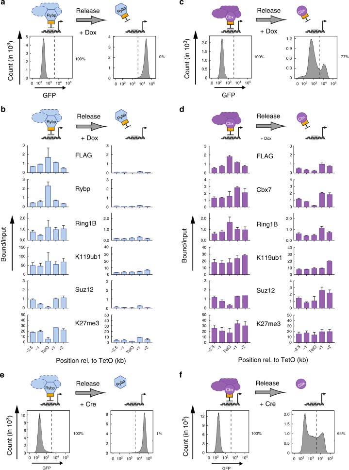
Fig. 2.
cPRC1 but not vPRC1 supports propagation of repressive chromatin. a, c Flow cytometry histograms relate GFP expression before and after release of TetR fusion protein recruitment in response to Dox treatment for 6 days. Percentages (%) indicate fraction of silenced cells. b, d ChIP qPCR analyses comparing relative enrichments of FLAG-TetR fusion proteins, PcG proteins and histone modifications before and after 6 days of Dox treatment. ChIP enrichments for H2AK119ub1 and H3K27me3 are normalized to negative control locus (IAP). Data are mean ± SD (error bars) of at least two independent experiments. Source data are provided as a Source Data file. e, f Flow cytometry histograms relate GFP expression before and 6 days after genetic release of Rybp and Cbx7 tethering by Cre-mediated deletion of the TetR DNA binding domain. Percentages (%) indicate fraction of silenced cells

