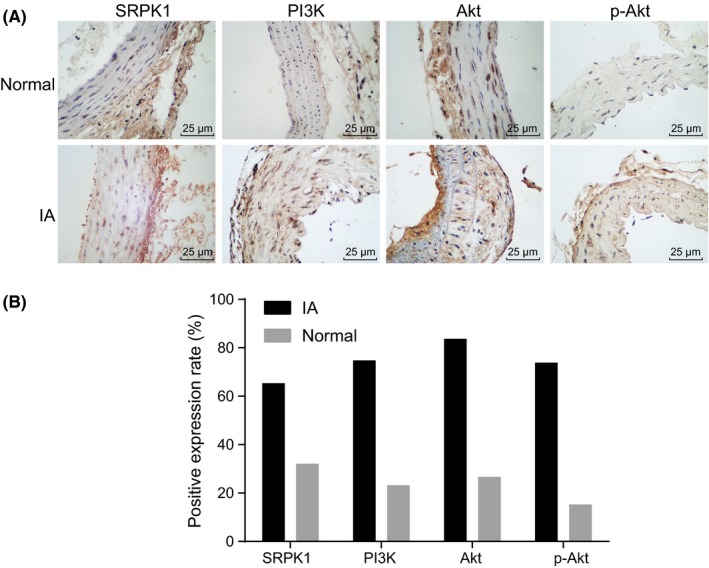
Figure 2.

Immunohistochemical staining analysis for the expression of SRPK1 and the PI3 K/Akt signaling pathway‐related factors (PI3 K, AKT, and p‐AKT) in the aneurysm wall of IA and normal vasculature. Panel A, immunohistochemical staining of SRPK1, PI3 K, Akt, and p‐Akt in the normal group and the IA group (400×). SRPK1 was mainly localized in the tunica media and tunica adventitia of the vascular wall, and in very little amounts in the tunica intima. PI3 K, Akt, and p‐Akt were mainly located in the tunica intima of vascular wall.; Panel B, quantitative analysis indicates expressions of SRPK1, PI3 K, Akt, and p‐Akt were up‐regulated in IA; *P < 0.05 vs the normal group; IA, intracranial aneurysm; SRPK1, SR protein‐specific kinases; PI3 K, phosphatidylinositol‐3 kinase
