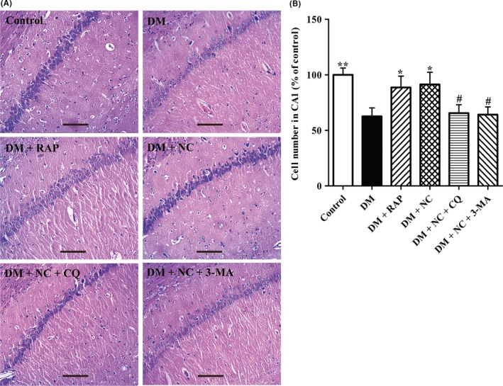
Figure 3.

Effect of NC on the histological features in the hippocampal CA1 region of DM rats induced by STZ. DM rats were treated with RAP (1 mg/kg/day, IP), NC (100 mg/kg/day, IP), NC (100 mg/kg/day, IP) + CQ (40 mg/kg/day, IP), NC (100 mg/kg/day, IP) + 3‐MA (1.5 mg/kg/day, IP) for 35 consecutive days. H&E staining was used to measure the changes in neuronal morphology in the hippocampal CA1 area. (A) Representative light micrographs of HE‐stained neuron cells in the hippocampal CA1 area of the rats. (B) Statistical analyses for the cell number in the hippocampal CA1 region of the rats. The data are expressed as mean ± SEM (n = 3 per group). *P < 0.05, **P < 0.01, compared with the DM group; # P < 0.05 compared with the DM + NC group. Scale bar, 100 μm
