
Figure 6.
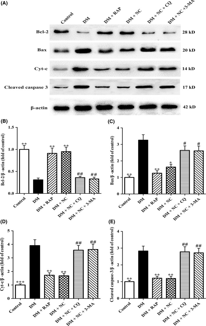
Effect of NC on apoptosis‐associated proteins Bcl‐2, Bax, Cyt‐c, and Caspase‐3 in the CA1 hippocampus of DM rats induced by STZ. Diabetic rats were treated with RAP (1 mg/kg/day, IP), NC (100 mg/kg/day, IP), NC (100 mg/kg/day, IP) + CQ (40 mg/kg/day, IP), NC (100 mg/kg/day, IP) + 3‐MA (1.5 mg/kg/day, IP) for 35 consecutive days. (A) Western blot analysis was performed to detect apoptosis‐associated proteins of Bcl‐2 (28 kD), Bax (20 kD), Cyt‐c (14 kD), and cleaved Caspase‐3 (17 kD) in the hippocampal CA1 region of rats. (B, C, D, and E) Densitometry analysis of Bcl‐2 (28 kD), Bax (20 kD), Cyt‐c (14 kD), and cleaved Caspase‐3 (17 kD) protein levels were performed by the NIH Image J software. β‐actin was used as control for protein loading. The data are expressed as mean ± SEM (n = 4 per group). *P < 0.05, **P < 0.01, ***P < 0.01, compared with the DM group; # P < 0.05, ## P < 0.05 compared with the DM + NC group