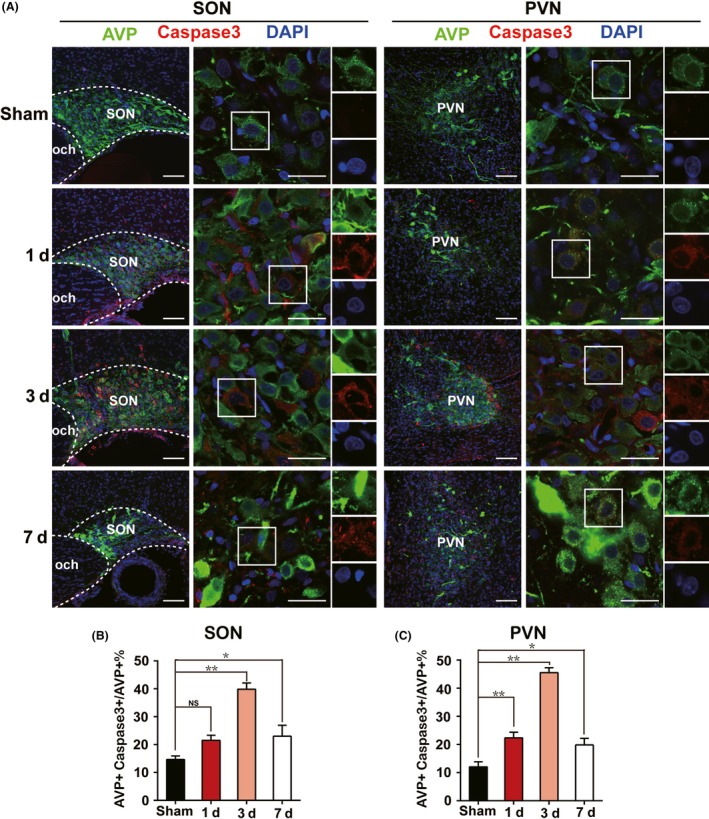
Figure 2.

Emerging of apoptotic AVP neurons in acute phase after PEL surgery. A, Immunofluorescent analysis of apoptotic AVP neurons of SON or PVN characterized by AVP+ Caspase3+ neurons at day 1 (N = 3), day 3 (N = 3), and day 7 (N = 3) after PEL surgery compared with sham‐operated rats (N = 3), respectively. B, Quantification of SON in A. C, Quantification of PVN in A; **P < 0.01 compared to sham‐operated rats, *P < 0.05 compared to sham‐operated rats; scale bars, 100 μm for low magnification image, 30 μm for high magnification image. AVP (green), Caspase3 (Red), DAPI (blue). AVP, arginine vasopressin; SON, supraoptic nucleus; PVN, paraventricular nucleus
