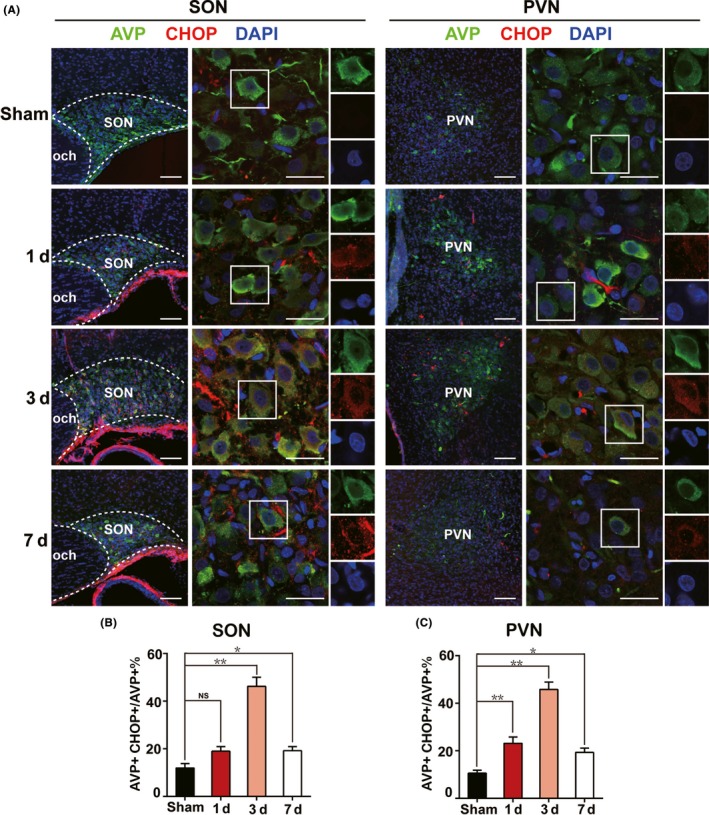
Figure 3.

Triggered ER stress in AVP neurons in acute phase after PEL surgery. A, Immunofluorescent images visualized that ER stress was triggered in AVP neurons of SON or PVN characterized by AVP+ CHOP+ neurons at day 1 (N = 3), day 3 (N = 3), and day 7 (N = 3) postsurgery in PEL and sham‐operated rats (N = 3), respectively. B, Quantification of SON in A. C, Quantification of PVN in A; **P < 0.01 compared to sham‐operated rats, *P < 0.05 compared to sham‐operated rats; scale bars, 100 μm for low magnification image, 30 μm for high magnification image. AVP (green), CHOP (Red), DAPI (blue). AVP, arginine vasopressin; SON, supraoptic nucleus; PVN, paraventricular nucleus
