Figure 6.

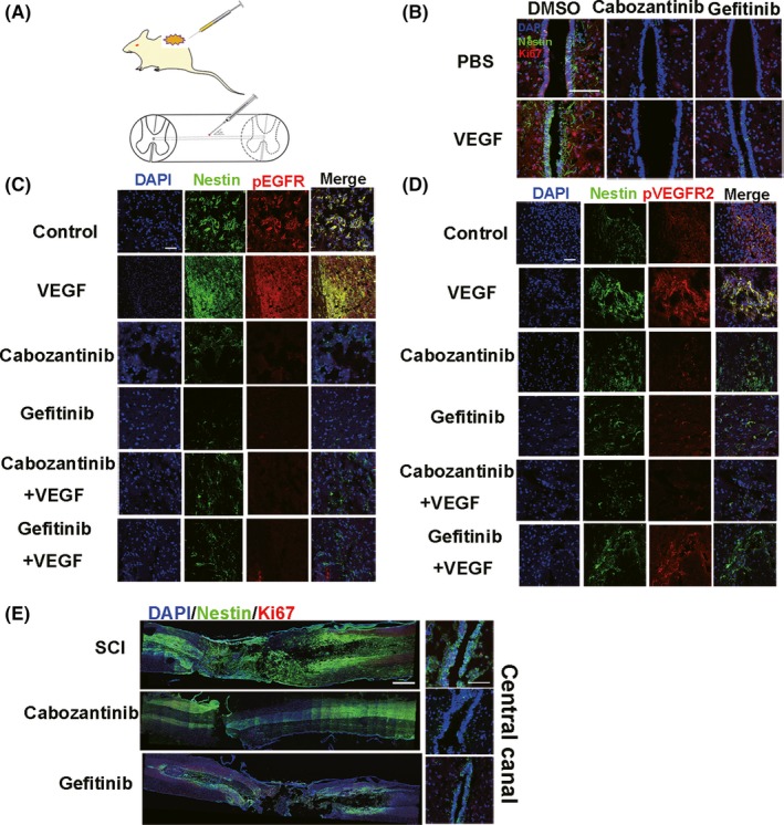
VEGF activates spinal cord NSCs through VEGFR2‐EGFR signaling on day 3 after injection in vivo (n = 5 rats/group). A, Model of the spinal cord intraspinal injection. The needle is about 25 gauge, at a 45° angle to the spinal cord (see Materials and Methods). The zigzag region indicates the injection site (T8‐T9). B, Injection of cabozantinib and gefitinib decreases the number of nestin+/Ki67+ spinal cord NSCs in the central canal. Injection of combination of PBS and DMSO as control. The results show that injection induced NSCs activation in central canal. Scale bar represents 200 µm. C, Nestin (green) and pEGFR (red) staining at the site of injection. Scale bar represents 100 µm. D, Nestin (green) and pVEGFR2 (red) staining at the site of injection. Scale bar represents 100 µm. A number of activated NSCs aggregate around the injection site (B‐D). E, Cabozantinib and gefitinib reduce nestin+/Ki67+ NSCs after SCI. Left, whole spinal section. Right, central canal; dpi, days postinjury. Nestin (green), Ki67 (red). Scale bar represents 500 µm in left and 200 µm in right panel
