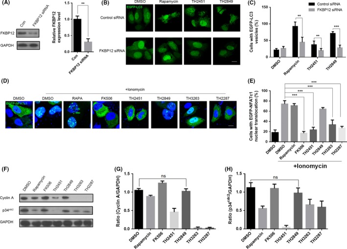
Figure 5.

TH2849 potentially induces autophagy by forming a complex with FKBP12 without interfering to the calcineurin/NFAT and IL2/p34cdc2/cyclin A signal pathway. (A) PC12 cells were pretreated with siRNA against FKBP12 for 48 h, and then, the protein and mRNA levels were examined. Representative images (B) and quantification (C) of PC12 cells with EGFP‐LC3 vesicles (autophagosomes) in the presence or absence of siRNA against FKBP12. PC12 cells co‐transfected with siRNA against FKBP12 and EGFP‐LC3 were treated with 1‰ DMSO, 1 μmol/L rapamycin, 10 μmol/L FK506, 1 μmol/L TH 2451 and 1 μmol/L TH 2849 for 24 h. (D) Representative images of distribution of EGFP‐NFATc1. The EGFP‐NFATc1 transfected MCF‐7 cells were treated with 2 μmol/L ionomycin or co‐treated with 1‰ DMSO, 1 μmol/L rapamycin, 1 μmol/L FK506, TH2451, TH 2849, TH3263 and TH2287 for 2 h as indicated. (E) Quantification of nucleus translocated EGFP‐NFATc1 cells in (D). (F) Representative Western blotting image of p34cdc2 and cyclin A levels. Factor‐deprived CTLL‐2 cells were cultured for 14 h in basic medium only, 50 units/mL IL‐2 with 1‰ DMSO, 1 μmol/L rapamycin, 1 μmol/L FK506, TH2451, TH 2849, TH3263, and TH2287 were added respectively during the last 1 hour. Quantification of relative p34cdc2 (G) and cyclin A (H) expression levels in (F). All the substances were dissolved in DMSO. Scale bars: 10 μm. Data are shown as the mean ± SEM *P < 0.05, **P < 0.01, ***P < 0.001 compared to DMSO control, n = 3.
