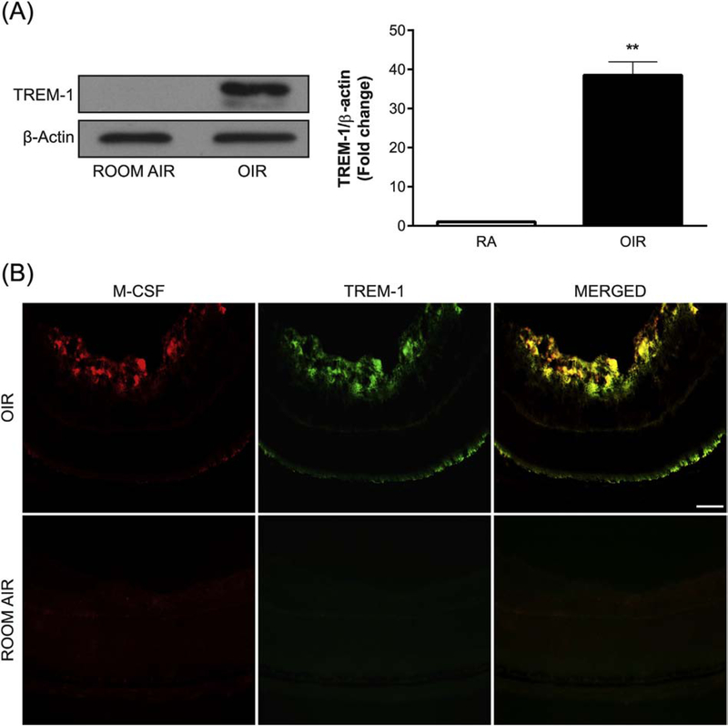
Fig. 1.
OIR induces TREM-1 and M-CSF expression. (A) A representative Western blot shows TREM-1 expression at P17 in the retinas of OIR mice but not of those kept in room air (RA). The membrane was probed for TREM-1 and then reprobed for β-actin. Values in the bar graphs represent the mean ± SEM, n = 5. **, p < 0.01 vs. RA mice. (B) Representative retinal cryosections from OIR and RA mice at P17 were immunolabeled with antibodies against M-CSF (red) and TREM-1 (green). TREM-1 and M-CSF are induced and largely colocalized in OIR. Scale bar = 20 μm. Five retinas were analyzed for each experimental group.

