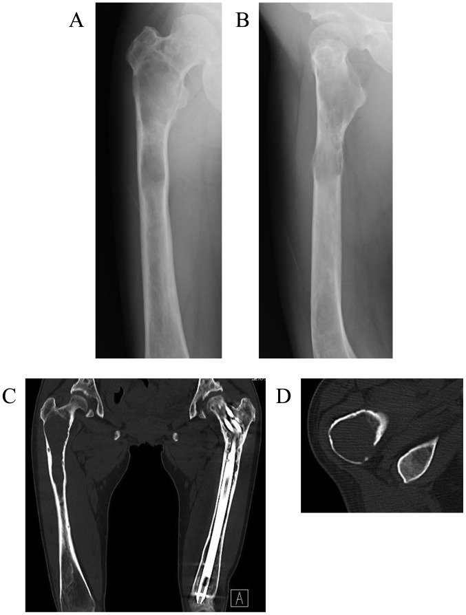
Figure 2.
Imaging findings when the same patient presented again 2 years later with a 5-month history of right thigh pain. Plain radiographs of the (A) anteroposterior and (B) lateral view and (C) plain coronal computed tomography (CT) revealed marked worsening in the changes of the right proximal femur compared with those from 2 years earlier, consisting of a central longitudinally medullary lytic lesion with expansion and thinning of the bone cortex. (D) Plain axial CT revealed slight disruption of the intertrochanteric posterior bone cortex, but no soft tissue mass or periosteal reaction were identified.

