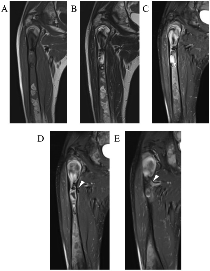
Figure 3.
Magnetic resonance imaging (MRI) findings of the patient at the same time as Fig. 2. Coronal MRI revealed an extended intramedullary hypointense lesion both on (A) T1- and (B) T2-weighted images. (C-E) Coronal fat-suppressed post-contrast T1-weighted MRI revealed heterogeneous enhancement around the intramedullary hypointense lesion, as well as several feeding arteries (arrowheads) arising from the deep femoral artery.

