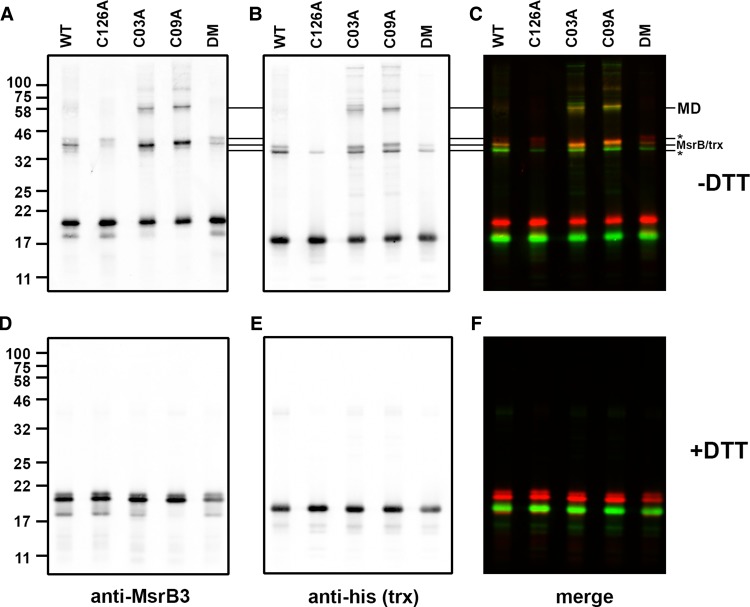
Figure 4. Formation of mixed disulfides between thioredoxin and MsrB3 cysteine mutants.
(A–F) The substrate-trapping mutant of thioredoxin was incubated with WT and various cysteine mutants of MsrB3 as indicated and separated by SDS–PAGE either in the absence (A–C) or presence (D–F) of DTT. Immunoblotting, scanning and image presentation is as described in Figure 3.

