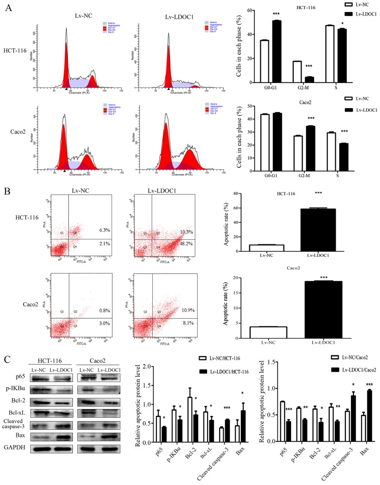
Figure 3.
Apoptosis rate and cell cycle phase distribution in the transfected cells were detected by flow cytometry, and the expression of apoptosis-associated proteins was analyzed by western blotting. (A) From the cell-cycle distributions, HCT116 (Lv-LDOC1) cells were significantly arrested in the G0/G1 phase and Caco2 (Lv-LDOC1) cells were significantly arrested in the G2/M phase. (B) The apoptosis rates of Lv-LDOC1 were increased, compared with Lv-NC. (C) Lv-LDOC1 increased cleaved caspase-3 and Bax levels and reduced p65, p-IKBα, Bcl-2 and Bcl-xl levels, compared with Lv-NC. *P<0.05, **P<0.01 and ***P<0.001 vs. Lv-NC. Bcl-2, B-cell lymphoma-2; Bcl-xl, B-cell lymphoma extra-large; Bax, Bcl-2-associated X protein; p-IKBα: phosphorylated inhibitor-κ-Bα; Lv-LDOC1, LDOC1-overexpressing cells; Lv-NC, negative control cells.

