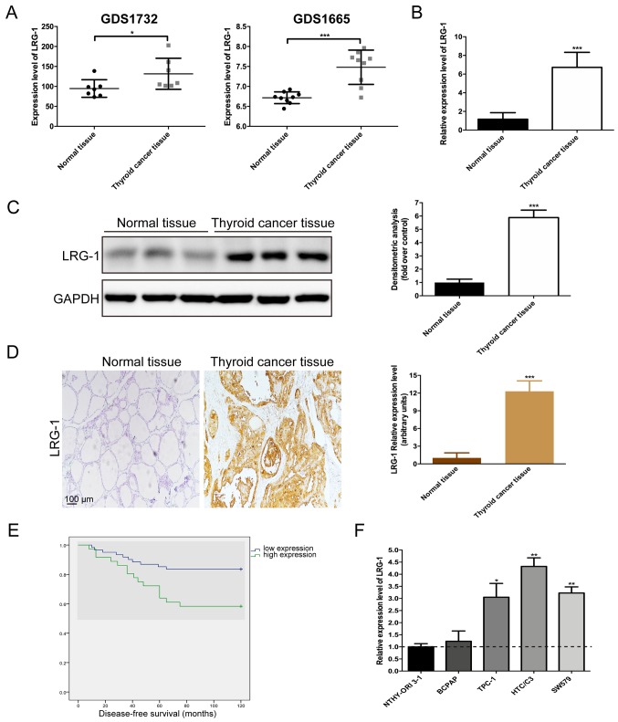
Figure 1.
LRG-1 is overexpressed in thyroid cancer tissues. (A) The mRNA expression of LRG-1 in thyroid cancer tissues and normal tissues based on the microarray data from GDS1732 (7 thyroid cancer tissues and 7 normal tissues) and GDS1665 (9 thyroid cancer tissues and 9 normal tissues). (B) The mRNA and (C) protein expression of LRG-1 in normal thyroid tissues (n=97) and thyroid cancer tissues (n=97). (D) Immunohistochemistry images of LRG-1 and the quantification of LRG-1 relative expression levels. Scale bars, 100 µm. (E) Disease-free survival analysis of thyroid cancer patients (n=97) with different LRG-1 expression levels. (F) Assessment of LRG-1 mRNA abundance in a normal thyroid cell line (NTHY-ORI3-1) and thyroid cancer cell lines (BCPAP, TPC-1, HTC/C3 and SW579) via qPCR. The horizontal dotted line represents LRG-1 mRNA levels in NTHY-ORI3-1 cells (black bar). Data are presented as the mean ± SD. *P<0.05; **P<0.01; ***P<0.001 vs. control (normal thyroid tissues or normal thyroid cell line). LRG-1, leucine-rich-alpha-2-glycoprotein 1.

