

Abstract
Minor vein ultrastructure and phloem loading were studied in leaves of the tulip tree (Liriodendron tulipifera; Magnoliaceae). Plasmodesmatal frequencies leading into minor vein companion cells are higher than in species known to load via the apoplast. However, these companion cells are not specialized as “intermediary cells” as they are in species in which the best evidence for symplastic phloem loading has been documented. Mesophyll cells plasmolyzed in 600 mm sorbitol, whereas sieve elements and companion cells did not plasmolyze even in 1.2 m sorbitol, indicating that solute accumulates in the phloem against a steep concentration gradient. Both [14C]sucrose and 14C-labeled photo-assimilate accumulated in the minor vein network, as demonstrated by autoradiography. [14C]sucrose accumulation was prevented by p-chloromercuribenzenesulfonic acid, an inhibitor of sucrose-proton cotransport from the apoplast. p-Chloromercuribenzenesulfonic acid largely, but not entirely, inhibited exudation of radiolabeled photoassimilate. The evidence is most consistent with the presence of an apoplastic component to phloem loading in this species, contrary to speculation that the more basal members of the angiosperms load by an entirely symplastic mechanism.
The role of plasmodesmata in phloem loading is enigmatic. Because solute movement through plasmodesmata is passive, photoassimilate should leak out of sieve elements (SEs) and companion cells (CCs) into symplastically-coupled cells and dissipate the gradient built up by active transport mechanisms.
In stems this is not a problem because the phloem is symplastically isolated (van Bel and Kempers, 1991; van Bel and van Rijen, 1994). In the minor veins of leaves, however, where phloem loading occurs, the situation is more complex. Indeed, in many species, plasmodesmatal frequencies are very high between minor vein CCs and adjacent phloem parenchyma (PP) or bundle sheath (BS) cells (Turgeon, 1996). By Gamalei's (1989) definition, these are “type- 1” plants. It has been suggested that the numerous plasmodesmata in type-1 species, rather than allowing solute leakage from CCs, are involved in symplastic loading (Gamalei, 1989; van Bel, 1993; Grusak et al., 1996; Turgeon, 1996). By the same reasoning, species with relatively few plasmodesmata at these interfaces (type-2 plants; Gamalei, 1989) are often assumed to load from the apoplast.
Classifying species into apoplastic and symplastic loading groups is the starting point for assessing evolutionary trends and ecological adaptiveness of different phloem loading strategies. For example, on the basis of comparative plasmodesmatal frequency data, it has been hypothesized that symplastic loading is the ancestral condition in angiosperms, and that apoplastic loading arose later in temperate and arid climates (Gamalei, 1991; van Bel and Gamalei, 1992).
However, if plants are to be classified as “symplastic loaders” or “apoplastic loaders” based on plasmodesmatal frequencies, it is important that this primary assumption be rigorously tested. The mechanism of loading from the apoplast (Suc-proton symport) is well established (Grusak et al., 1996; Komor et al., 1996; Rentsch and Frommer, 1996), but the mechanism(s) of symplastic loading are more obscure. It is unclear that a plant can be assumed to be a symplastic loader based on plasmodesmatal frequency data alone.
Putative symplastic-loading species (type 1) fall into at least two groups based on the sugars they transport and the anatomy of their veins (Turgeon, 1995). Those in the smaller, but better studied, of these groups translocate high proportions of raffinose and stachyose in addition to Suc. The cucurbits are an example. Their minor vein CCs appear to be structurally adapted to symplastic transfer from the BS and are known as “intermediary cells.” A “polymer trap” mechanism of symplastic loading, consistent with thermodynamic principles, has been proposed for plants with intermediary cells (Turgeon, 1996).
It is unfortunate that very little work has been done on the second, larger group of putative symplastic loaders. Whereas they have numerous plasmodesmata leading from the mesophyll into the minor vein phloem, they translocate Suc with only small amounts of raffinose and stachyose and therefore seem not to be candidates for polymer trapping. Many of these plants are trees (Gamalei, 1991). We chose to study the tulip tree (Liriodendron tulipifera; Magnoliaceae) as a representative of this group. Because the tulip tree occupies a relatively basal phylogenetic position (Nandi et al., 1998; Soltis et al., 1998), information on this species should provide insight into the evolution of phloem loading.
RESULTS
Minor Vein Structure
Analysis of the architecture of minor veins can provide insight into the routes and mechanisms of assimilate loading. In the tulip tree, SEs and CCs form one or two clusters in the center of the vein (Fig. 1). These complexes are in close association with PP cells. In the smallest minor veins, with only one or two SEs, the CCs come into direct contact with both PP cells and BS cells. However, in larger minor veins, PP cells ring the SE-CC complexes so that direct contact with the BS becomes less frequent.
Figure 1.

Minor vein in a tulip tree leaf. The vein is composed of a tracheary element (T), four SEs (S), and four CCs (C) surrounded by parenchyma cells (P). The vein is encircled by BS cells (B) with dense osmiophilic bodies in the vacuoles. The secondary wall thickening of the tracheary element is in the upper right corner of the cell and is not shown. Bar = 2 μm.
In transverse sections, there is one CC and approximately two PP cells for each SE. PP cells have the greatest mean cross-sectional area (21.4 μm2; se = 1.13 μm2; n = 170), followed by CCs (11.4 μm2; se = 0.83 μm2; n = 76) and SEs (3.11 μm2; se = 0.21 μm2; n = 76). In the largest minor veins, PP cells near the center of the vein are smaller than those closest to the BS, which appear to inter-grade with BS cells in some instances.
Numbers of interfaces and total interface lengths are given in Table I for veins with four SEs, the size of vein most frequently encountered. The longest total interface per vein is between BS and PP cells. Contact between adjacent CCs is less extensive than between CCs and PP cells.
Table I.
Interfaces and plasmodesmata (pd) in tulip tree leaves
| Interface | Per 4-SE
Vein
|
Pd μm−2 | Branching Ratio | |
|---|---|---|---|---|
| No. of interfaces | Total interface length (μm) | |||
| PP/CC | 9.00 ± 0.85 | 20.6 ± 2.21 | 3.65 | 1.57* |
| BS/PP | 11.7 ± 1.67 | 42.9 ± 6.82 | 3.05 | 1.07 |
| BS/CC | 1.83 ± 0.87 | 5.27 ± 2.61 | 3.31 | 0.65 |
| CC/SE | 6.67 ± 0.23 | 12.2 ± 0.75 | 2.84 | 14.3* |
| PP/SE | 5.67 ± 1.19 | 7.75 ± 2.06 | 1.32 | ID |
| CC/CC | 0.67 ± 0.23 | 1.10 ± 0.44 | 3.14 | NA |
| PP/PP | 10.0 ± 3.62 | 24.8 ± 8.25 | 6.29 | NA |
| BS/SE | 0.5 ± 0.54 | 0.67 ± 0.73 | Rare | ID |
| SE/SE | 1.5 ± 0.26 | 2.0 ± 0.22 | 2.54 | NA |
| MC/MC | NA | NA | 3.09 | NA |
Numbers of interfaces, and total length of each interface type, were measured in transverse sections of veins with four sieve elements, the most common size sampled. Pd μm−2 of interface was calculated from data on veins of all sizes. Pd were counted only if they traversed at least halfway across the common wall between cells. When pd on the two sides of the wall were disconnected, they were each considered to be a single plasmodesma. The branching ratio was calculated as the ratio of pd on the opposing sides of a common wall. CC, Companion cell; PP, phloem parenchyma cell; SE, sieve element; MC, mesophyll cell; ID, insufficient data; NA, not applicable; ± se; *, statistically different from 1.0 at the 0.5 confidence level using a paired t test.
PP cells and CCs are similar in internal structure (Fig. 1). They are vacuolate with relatively dense cytoplasm. Both cell types have chloroplasts, which are considerably smaller than the chloroplasts of BS or mesophyll cells. Starch is present in the chloroplasts of PP cells, but rare in CCs. CCs can often be identified by their spatial relationship to adjacent SEs, but if this is not the case, the distinction between CCs and PP cells is frequently difficult to discern. PP cells adjacent to the BS sometimes contain osmiophilic bodies as do BS and mesophyll cells.
Plasmodesmata and the Route of Phloem Loading
Plasmodesmatal frequencies at different interfaces in the minor vein are relatively similar in the tulip tree leaves (Table I). They are most frequently found between adjacent PP cells and are least common between PP cells and SEs. Similar numbers of plasmodesmata are found at the interfaces between mesophyll cells.
Since frequencies are similar, total numbers of plasmodesmata at the different interfaces are determined primarily by interface lengths. This is illustrated in Figure 2 in which the numbers of plasmodesmata at the different interfaces are summed from all of the 21 veins studied. Whereas the frequency of plasmodesmata between BS cells and either PP cells or CCs is similar (Table I), there are far more total plasmodesmata at the former interface than at the latter (Fig. 2), indicating that PP cells may be the major conduit of photoassimilate into the vein. Note also that there are few plasmodesmata linking adjacent CCs or adjacent SEs, i.e. individual SE-CC complexes in the vein are for the most part symplastically isolated from one another.
Figure 2.

Total number of plasmodesmata recorded at each interface type in transverse sections of 21 tulip tree minor veins. Most plasmodesmata are found in the walls of PP cells.
Plasmodesmata between PP cells (Fig. 3A) and between CCs or BS cells (Fig. 3B) are branched on both sides from a central median cavity. At the site of the plasmodesmata, the wall is noticeably thickened. The plasmodesmal branches appear to be approximately the same width on both sides of the wall. In favorable sections, desmotubules can be seen; however, the substructure of plasmodesmata was not analyzed in detail in this study. Branching frequencies were obtained by counting branches on each side of the common walls between different cell types (Table I). Plasmodesmata branches between PP cells and CCs are more common on the PP side, although the difference is not great. As has often been noted in other species, plasmodesmata between CCs and SEs are much more common on the CC side.
Figure 3.

Plasmodesmata between PP cells (A) and between PP cell (at right) and CC (B) in tulip tree minor veins.
Plasmolysis and Suc Concentration
To determine the relative solute concentrations of cells along the phloem loading pathway, leaf tissue samples were plasmolyzed in sorbitol solution, fixed, and examined in the electron microscope (Beebe and Evert, 1992). Mesophyll cells were unplasmolyzed in 500 mm sorbitol but uniformly plasmolyzed in 600 mm sorbitol. There was rarely evidence of plasmolysis in SEs and CCs at any sorbitol concentration, including 1.2 m, the highest concentration used. PP cells were more resistant than mesophyll cells to plasmolysis; at 600 mm sorbitol they were unplasmolyzed, although at 700 mm and above, plasmolysis was generally evident. The concentration of Suc in mature leaves was 2.6 nmol mm−2.
14C Accumulation in Veins
To determine if photoassimilate accumulates in the minor vein network, we exposed leaves to 14CO2 for 5 min, followed by a chase in normal atmosphere. The leaf tissue was then flash-frozen, lyophilized, pressed flat, and autoradiographed with x-ray film. In our initial experiments, no vein images were obtained. Reasoning that the cuticle may be too thick to permit resolution of the veins, we repeated the experiments, removing the lower epidermis and most of the spongy mesophyll prior to freezing the tissue. In these preparations, vein images were not obtained if the tissue was frozen immediately after the 5-min exposure to 14CO2 (not shown), but they were consistently seen after a chase period of 55 or 85 min (Fig. 4).
Figure 4.
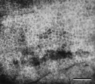
Autoradiograph of tissue from an attached tulip tree leaf exposed to 14CO2 for 5 min and to normal atmosphere for 85 min. The lower epidermis and most of the spongy mesophyll was removed before freezing and lyophilizing the tissue. The minor veins accumulated radiolabeled photo-assimilate. The x-ray film was used as the photographic negative; therefore, radiolabel appears white. Bar = 1.5 mm.
Essentially the same results were obtained when excised leaf tissue was floated on [14C]Suc solution (Fig. 5, A and B). Images of the vein network, indicating accumulation of radiolabel, were not obtained if leaf tissue was abraded by standard procedures using carborundum or sandpaper. However, when the lower epidermis and most of the spongy mesophyll was removed, accumulation of radiolabel in the veins could be seen (Fig. 5A). Contrast was very low; veins images were only seen in those areas of the tissue where the razor blade passed just below the vein network. However, the results were consistent in all four replicate experiments.
Figure 5.

Autoradiographs of tulip tree (A and B) and C. blumei (C and D) leaf tissue exposed to [14C]Suc. The lower epidermis and most of the spongy mesophyll was removed with a razor blade and the tissue was floated, cut surface down, on 1 mm [14C]Suc for 30 min with (B and D) or without (A and C) the addition of 5 mm PCMBS to the solution. The tissue was then washed for 30 min, frozen, and lyophilized. Vein images are visible in both the tulip tree (A) and C. blumei (C) due to accumulation of radiolabel. PCMBS prevented accumulation of [14C]Suc in veins of the tulip tree (B) but not in C. blumei (D). The x-ray films were exposed for 3 d in A, C, and D and for 9 d in B. All images are at the same magnification; bar in D = 1.5 mm.
p-Chloromercuribenzenesulfonic acid (PCMBS; 5 mm) considerably reduced the amount of [14C]Suc taken up by the tissue. To compensate for the reduced radiolabel, x-ray films were exposed for a range of periods to achieve similar degrees of film density. No matter how long the films were exposed, no accumulation of [14C]Suc by veins was seen in tissue treated with PCMBS (Fig. 5B).
Vein loading images were more pronounced in autoradiographs of Coleus blumei leaf tissue treated in the same fashion (Fig. 5C), at least partly due to the wide vein spacing. PCMBS did not inhibit phloem loading in C. blumei (Fig. 5D), as previously shown (Weisberg et al., 1988).
Effects of PCMBS on Exudation
Exudation of 14C-labeled photosynthate from excised leaves into a solution of EDTA (King and Zeevaart, 1974) was measured to analyze the effect of PCMBS on phloem-loading and long-distance translocation. Approximately 0.17 mL of PCMBS solution entered each leaf as determined by weighing the uptake vials before and after the 30-min transpiration period. Safannin O solution (0.1%; w/v) permeated the minor vein system within 15 min when administered in the same way (data not shown).
Following the labeling period, leaves exuded radioactivity for more than 20 h (Fig. 6A). Cumulative exudation was approximately logarithmic with time for the first 6 or 8 h (Fig. 6B). PCMBS (0.5 mm) inhibited exudation to a considerable degree, but not entirely, when administered via the transpiration stream (Fig. 6, A and B). At the end of the 20-h experiments, PCMBS-treated leaves had exuded approximately 20% of the label exuded by control leaves. In some experiments a small amount of damage to the lamina was apparent as watery lesions in the PCMBS-treated leaves, especially at the extremities of the blade. Treatment with 0.5 mm PCMBS did not inhibit photosynthetic uptake of 14CO2 (Fig. 7). Leaves exposed to 1.0 mm PCMBS by the same method were extensively damaged by the treatment and exudation was almost entirely eliminated (not shown).
Figure 6.

Exudation of 14C-labeled photo-assimilate into EDTA solution from detached tulip tree leaves. Water (□) or PCMBS (0.5 mm; ▪) was administered to leaves via the transpiration stream prior to photosynthesis in 14CO2 and measurement of photosynthesis (see Fig. 7). The cut ends of petioles (six per sample) were immersed in EDTA (20 mm) solution to prevent callose formation, and the exudate was periodically counted. Data are plotted on a cumulative basis on linear (A) and logarithmic (B) scales. Bars = se (n = 4).
Figure 7.

Photosynthetic incorporation of 14CO2 by detached tulip tree leaves used in the experiments reported in Figure 6. Following administration of water or PCMBS (0.5 mm) by the transpiration stream, the leaves were exposed to 14CO2 and radiolabel was measured with a β-scintillator tube pressed against the leaf surface. Bars = se (n = 24).
DISCUSSION
One of our primary objectives was to determine if phloem loading occurs in the tulip tree. This is a relevant issue since photoassimilate accumulation against a concentration gradient has been demonstrated in species with intermediary cells (Turgeon and Gowan, 1990; Haritatos et al., 1996) but not in type-1 species without this distinctive cell type. To the contrary, several lines of evidence indicate that Suc is not loaded against a concentration gradient into the minor vein phloem in willow (Salicaceae) (Turgeon and Medville, 1998). In addition, the plasmolysis data on Populus deltoides (Salicaceae) in the paper by Russin and Evert (1985) can be interpreted in the same way (Orlich et al., 1998; Turgeon and Medville, 1998). Thus, it is reasonable to ask if the absence of phloem loading is a common or even a universal feature of other species in this category of type-1 plants.
The evidence presented here indicates that this is not the case. In the tulip tree, the osmotic potential of minor vein phloem, estimated by plasmolysis, is much higher than that of the mesophyll, demonstrating the presence of a strong solute gradient as shown in many other species with documented phloem loading (Turgeon and Medville, 1998). In willow, the concentration of Suc in leaves is very high (16.0 nmol mm−2; Turgeon and Medville, 1998) presumably to generate a diffusion gradient from the mesophyll to the veins. In contrast, the concentration of Suc in the tulip tree leaves (2.6 nmol mm−2) is less than one-sixth this value.
The next question to be addressed is whether loading is apoplastic or symplastic. The pathway(s) that assimilates take from the mesophyll to the phloem are difficult to resolve. For example, loading has been extensively studied in Ricinus communis cotyledons, yet only recently has it been found that there is a measurable symplastic component in addition to the extensively documented apoplastic transfer (Orlich et al., 1998).
One approach to distinguishing between apoplastic and symplastic loading is to analyze minor vein anatomy and plasmodesmatal frequencies. Species for which the best evidence for apoplastic loading has been derived are tobacco, sugar beet, Vicia faba, and R. communis (Grusak et al., 1996; Komor et al., 1996; Rentsch and Frommer, 1996). The first three fall into Gamalei's type-2 category with low symplastic continuity into the minor vein phloem (Gamalei, 1989). R. communis is in an intermediate category (type 1–2a; Gamalei, 1989) between type 1 and type 2. As discussed above, R. communis loads via the apoplast and may also have a symplastic component to loading (Orlich et al., 1998).
There is not enough known about symplastic loading to be able to predict its occurrence on the basis of plasmodesmatal frequency data alone. The best evidence for symplastic loading comes from species with intermediary cells (Turgeon, 1996), specialized CCs in the minor vein phloem. Intermediary cells are densely cytoplasmic with small vacuoles and rudimentary plastids. They always abut the BS to which they are joined by extensive fields of branched plasmodesmata. Plasmodesmatal branches are narrower and more numerous on the intermediary cell side than on the BS side (Fisher, 1986). Plants with intermediary cells translocate a substantial proportion of their carbohydrate as raffinose and stachyose (Turgeon, 1996).
From the results of Zimmermann and Ziegler (1975), indicating that the tulip tree translocates only traces of raffinose and stachyose, we expected to find that this species does not have intermediary cells. This proved to be true. The CCs in the tulip tree minor veins are often separated from the BS by PP cells, and they do not have the characteristically dense cytoplasm or small vacuoles of intermediary cells. Furthermore, the plasmodesmata leading into them from BS cells or PP cells appear to be branched more on the PP side than on the CC side, and the branches are all of approximately the same width.
The one characteristic of these CCs that may indicate a symplastic mode of loading is high plasmodesmatal frequency. There is some disparity in this regard between our values for the tulip tree (3.65 plasmodesmata μm−2 between PP and CCs) and those of Gamalei (1991; 23.62 μm−2). This is partly due to a difference in methodology; we correct our data for section thickness, which reduces the value by a factor of approximately 3.4. Neglecting this correction brings our figure to 12. 4 μm−2, which is still only half that reported by Gamalei. Whatever the reason for this discrepancy, the tulip tree is still at the lower end of the range of type-1 plants in terms of plasmodesmatal frequency according to our data or those of Gamalei (1991).
The distinction between Gamalei's type-1 and type-2 plants is based on frequencies alone and is arbitrary. Therefore, there does not appear to be a compelling reason to postulate, on this basis, that loading in the tulip tree is symplastic when loading in R. communis is known to be largely, if not entirely, apoplastic. Although it may seem contrary to expectations for a plant that loads apoplastically to have many plasmodesmata at the interface with CCs, it is possible that the size exclusion limit at this interface is restricted or that the pores close in response to differences in turgor pressure between the two joined cell types (Oparka and Prior, 1992; see below). Either being the case, leakage would be minimal, and the frequency data may be largely or entirely irrelevant.
Another important consideration when analyzing solute transfer is that it is more meaningful to estimate plasmodesmatal numbers leading from one cell type to another than to rely on frequency data alone (Fisher, 1990); in other words, frequencies should be multiplied by interface lengths (or areas). On this basis, intermediary cells are also distinct. For example, in C. blumei, plasmodesmata at the BS-CC interface are more numerous than at any other interface in the leaf (see compiled data and plasmodesmograms of Botha and van Bel, 1992). This is not true in any of the other species analyzed by Botha and van Bel (1992), and it is not true of the tulip tree (Table I).
A second experimental approach used to distinguish between symplastic and apoplastic pathways is to analyze PCMBS sensitivity (Giaquinta 1976; van Bel et al., 1993; Flora and Madore, 1996; Weig and Komor, 1996). PCMBS blocks the action of Suc-proton symporter proteins located on the plasma membrane but does not inhibit symplastic transport (Turgeon, 1996). The effect is seen in macro-autoradiographs in which PCMBS eliminates the images of radiolabeled veins when leaf tissues are exposed to 14CO2 or are incubated in exogenous [14C]Suc solution. PCMBS effectively prevents accumulation of [14C]Suc from the apoplast in the tulip tree as demonstrated by the autoradiographic experiments reported here. The contrast in autoradiographic images of veins in the tulip tree is very low following exposure of leaf tissue to [14C]Suc. This could be due to limited uptake of exogenous label by the veins or to especially high rates of uptake into the mesophyll (van Bel et al., 1993).
Eschrich and Fromm (1994) did not detect vein images in autoradiographs in a number of plants, including some type-1 species without intermediary cells, following exposure to [14C]Suc. They concluded that photo-assimilate is not loaded via the apoplast in these plants. Unfortunately, the method of leaf abrasion used in their study was not described. As discussed above, it is important to remove the epidermis of leaf tissue from woody species in autoradiography experiments; standard abrasion techniques with carborundum, or even sandpaper, are not sufficient. Even with these treatments, images of veins are often weak.
Consistent with an apoplastic mechanism of phloem loading in the tulip tree, PCMBS also inhibited exudation of 14C-labeled photoassimilate from the cut ends of petioles into an EDTA solution (King and Zeevaart, 1974). Inhibition was apparent at 0.5 mm PCMBS; however, the effective concentration at which a substance is delivered to the minor venation via the transpiration stream is difficult to determine since evaporative loss of water concentrates the solution in the blade. Assuming that apoplastic water volume constitutes 11% of leaf fresh weight (Speer and Kaiser, 1991), the amount of PCMBS solution taken up (0.17 mL) by a single tulip tree leaf (2.4 g) is 64% of apoplastic water content. However, this probably underestimates the amount of PCMBS that comes in contact with the phloem as the solution is carried into the lamina in the directly adjacent xylem. The effectiveness of this delivery system is apparent in the damage caused by the inhibitor at concentrations that have no apparent damaging effect when leaf discs are floated on solutions. Damage to leaf tissues by transpired PCMBS was also noted in C. blumei leaves (Turgeon and Gowan, 1990).
PCMBS inhibition of exudation was not complete. Even when lamina tissue exhibited symptoms of damage by the inhibitor, some exudation, amounting to approximately 20% that of controls, continued. This is in contrast to the almost complete inhibition of exudation into EDTA solution in V. faba (Flora and Madore, 1996) and Pisum sativum (van Bel et al., 1993). These species have transfer cell ingrowths in the minor vein phloem and apparently load exclusively via the apoplast (Bourquin et al., 1990; Wimmers and Turgeon, 1991). The data on the tulip tree therefore indicate that there is a minor, PCMBS-insensitive pathway from the mesophyll into the phloem. The most likely avenue for this photo-assimilate movement is through plasmodesmata.
The most straightforward interpretation of these results is that there are two concurrently operating mechanisms of phloem loading in the tulip tree leaves, one apoplastic, the other symplastic. It may be that proportionate flux by the two pathways is not constant but changes under different physiological conditions, at different times of day, or in different seasons. No systematic study of these parameters was conducted. It is also possible that symplastic loading has no normal physiological role, but is simply induced by PCMBS treatment. If the plasmodesmata linking CCs with PP and BS cells are closed by the turgor pressure difference between these cell types (Oparka and Prior, 1992), they may open when PCMBS is administered; this could occur if Suc is transported away and is not replaced by the proton co-transport system, thus lowering turgor pressure in the minor vein phloem.
The finding that phloem loading in the tulip tree is sensitive to PCMBS is not consistent with the argument that all type-1 species load via the symplast. This conclusion has been based almost entirely on plasmodesmatal frequency data obtained from plants without intermediary cells and for which no detailed, compelling mechanism of symplastic loading has been put forward. It is reasonable, considering the data at hand, to re-evaluate the conclusion that all species with extensive symplastic connection to minor vein CCs load via the symplast. This being the case, the concept that symplastic phloem loading is the ancestral condition in angiosperm evolution should also be viewed with caution.
MATERIALS AND METHODS
Plant Material
Experiments were conducted on fully expanded leaves taken from mature, open-grown tulip trees (Liriodendron tulipifera; Magnoliaceae) on the Cornell University campus. Replicate experiments were conducted over three growing seasons.
Electron Microscopy
Pieces of lamina approximately 1.5 mm long on a side were fixed in 4% (w/v) glutaraldehyde in 100 mm sodium cacodylate buffer, pH 7.2, for 4 h at 4°C. The samples were post fixed in 1% (w/v) osmium tetroxide in the same buffer at 4°C overnight, dehydrated in ethanol, and embedded in Spurr resin. Plasmodesmatal counts and morphometric measurements were made on electron micrographs printed at 10,000× magnification or higher. Only those plasmodesmata extending at least halfway across the common wall between contiguous cells were counted. In the case of branched plasmodesmata, each branch was counted as one plasmodesma. Where branching was unequal in a complex, the numbers of branches on the two sides were averaged. The frequency of plasmodesmata per square micrometer was calculated by the method of Gunning described in the open discussion following the paper of Robards (1976).
Plasmolysis and Suc Concentration
Leaves were harvested and brought to the laboratory within 5 min. Details of the plasmolysis procedure have been described (Turgeon and Medville, 1998). Briefly, lamina pieces were excised under buffer, floated at room temperature on one of a series of sorbitol solutions ranging from 200 mm to 1.2 m, fixed in glutaraldehyde at the same osmolarity, post-fixed in osmium tetroxide, and embedded in Spurr resin for electron microscopy.
Leaf Suc concentration was measured by extracting tissue in methanol:chloroform:water (12:5:3; v/v), adding additional water to separate the aqueous phase, passing the aqueous phase through ion-exchange membranes, and analyzing sugars in the neutral fraction by HPLC. Procedures have previously been described in detail (Turgeon et al., 1993).
Autoradiography
Accumulation of radiolabeled photoassimilate in leaves was studied by enclosing attached leaves in full sun in a plastic bag and exposing them to 14CO2 (0.5 MBq), generated in the barrel of a syringe (Turgeon and Medville, 1998). After 5 min the bag was removed and 25 to 85 min later the leaves were removed from the tree and the lower epidermis and much of the mesophyll was quickly removed with a razor blade. The tissue was frozen in powdered dry ice, lyophilized in a −30°C chamber, pressed thin between steel plates in a large vice, and pressed, lower side down, against x-ray film (Hyperfilm-βmax, Amersham).
To analyze uptake of Suc from the apoplast, the lower epidermis and much of the spongy mesophyll was removed from segments of leaf tissue with a razor blade. The tissue was then floated, cut surface down on a solution of [14C]Suc (1.0 mm; 37 kBq mL−1) in 2(N-morpholino) ethanesulfonic acid (MES) buffer (20 mm MES plus 2.0 mm CaCl2, pH 5.5), with or without the addition of 5 mm PCMBS for 30 min followed by three 10-min washes in the same buffer. Both the initial uptake and washes were at room temperature. The tissue was frozen in powdered dry ice, lyophilized, pressed flat, and autoradiographed as described above.
Phloem Exudate
Shoots with several mature leaves were removed from trees, recut under water, and brought to the laboratory within 5 min. Petioles were cut under the surface of water, and leaves were distributed to vials (six leaves per vial) with the cut ends of the petioles immersed in either water or PCMBS solution. The leaves were placed under water-filtered lights (metal halide; 350 μmol photons m−2 s−1) for 30 min to allow transpiration of the water or PCMBS solution into the lamina. The amount of uptake was measured by weighing the vials. At the end of the uptake period the leaves were transferred to vials containing water and exposed to 14CO2 (1.7 MBq) for 1 h in a 16.9-L photosynthesis chamber under the same light conditions. At the end of the labeling period the amount of radiolabel incorporation was measured by placing a β-scintillator (model 44–1; Ludlum Measurements, Stillwater, TX) against the surface of each leaf in two nonoverlapping positions on opposite sides of the midrib. With the petioles still immersed in water, leaves were transferred to the dark for 45 min to close the stomata and prevent further transpiration. The petioles were then recut under water to a standard length of 4 cm, and the leaves were immediately transferred to plastic tubes containing 1.0 mL of EDTA solution (20 mm disodium EDTA, pH 7.0 with KOH) to prevent callose formation at the cut surface (King and Zeevaart, 1974). The leaves were placed in a dark, humid chamber to reduce transpiration; this inhibited reflux of exuded label and uptake of EDTA, which is toxic. At intervals, the EDTA solution was entirely removed for scintillation counting and replaced. Counts were adjusted for the small amount (approximately 30 μL h−1) of solution transpired (Araki et al., 1997).
Footnotes
This work was supported by the National Science Foundation (grant no. IBN–9603152 to R.T.).
LITERATURE CITED
- Araki T, Kitano M, Eguchi H. Evaluation of photoassimilate flux through a tomato pedicel. Biotronics. 1997;26:21–29. [Google Scholar]
- Beebe DU, Evert RF. Photoassimilate pathways(s) and phloem loading in the leaf of Moricandia arvensis (L.) DC. (Brassicaceae) Int J Plant Sci. 1992;153:61–77. [Google Scholar]
- Botha CEJ, van Bel AJE. Quantification of symplastic continuity as visualized by plasmodesmograms: diagnostic value for phloem-loading pathways. Planta. 1992;187:359–366. doi: 10.1007/BF00195659. [DOI] [PubMed] [Google Scholar]
- Bourquin S, Bonnemain J-L, Delrot S. Inhibition of loading of 14C assimilates by p-chloromercuribenzenesulfonic acid: localization of the apoplastic pathway in Vicia faba. Plant Physiol. 1990;92:97–102. doi: 10.1104/pp.92.1.97. [DOI] [PMC free article] [PubMed] [Google Scholar]
- Eschrich W, Fromm J. Evidence for two pathways of phloem loading. Physiol Plant. 1994;90:699–707. [Google Scholar]
- Fisher DG. Ultrastructure, plasmodesmatal frequency, and solute concentration in green areas of variegated Coleus blumei Benth. leaves. Planta. 1986;169:141–152. doi: 10.1007/BF00392308. [DOI] [PubMed] [Google Scholar]
- Fisher DG. Distribution of plasmodesmata in leaves: a comparison of Cananga odorata with other species using different measures of plasmodesmatal frequency. In: Robards AW, Lucas WJ, Pitts JD, Jongsma HJ, Spray DC, editors. Parallels in Cell to Cell Junctions in Plants and Animals. Berlin: Springer-Verlag; 1990. pp. 199–221. [Google Scholar]
- Flora LL, Madore MA. Significance of minor-vein anatomy to carbohydrate transport. Planta. 1996;198:171–178. [Google Scholar]
- Gamalei Y. Structure and function of leaf minor veins in trees and herbs: a taxonomic review. Trees. 1989;3:96–110. [Google Scholar]
- Gamalei Y. Phloem loading and its development related to plant evolution from trees to herbs. Trees. 1991;5:50–64. [Google Scholar]
- Giaquinta R. Evidence for phloem loading from the apoplast: chemical modification of membrane sulfhydryl groups. Plant Physiol. 1976;57:872–875. doi: 10.1104/pp.57.6.872. [DOI] [PMC free article] [PubMed] [Google Scholar]
- Grusak MA, Beebe DU, Turgeon R. Phloem loading. In: Zamski E, Schaffer AA, editors. Photoassimilate Distribution in Plants and Crops: Source-Sink Relationships. New York: Marcel Dekker; 1996. pp. 220–227. [Google Scholar]
- Haritatos E, Keller F, Turgeon R. Raffinose oligosaccharide concentrations measured in individual cell and tissue types in Cucumis melo L. leaves: implications for phloem loading. Planta. 1996;198:614–622. doi: 10.1007/BF00262649. [DOI] [PubMed] [Google Scholar]
- King RW, Zeevaart JAD. Enhancement of phloem exudation from cut petioles by chelating agents. Plant Physiol. 1974;53:96–103. doi: 10.1104/pp.53.1.96. [DOI] [PMC free article] [PubMed] [Google Scholar]
- Komor E, Orlich G, Alfons W, Koeckenberger W. Phloem loading-not metaphysical, only complex: towards a unified model of phloem loading. J Exp Bot. 1996;47:1155–1164. doi: 10.1093/jxb/47.Special_Issue.1155. [DOI] [PubMed] [Google Scholar]
- Nandi OI, Chase MW, Endress PK. A combined cladistic analysis of angiosperms using rbcl and non-molecular data sets. Ann Missouri Bot Garden. 1998;85:137–212. [Google Scholar]
- Oparka KJ, Prior DAM. Direct evidence for pressure-generated closure of plasmodesmata. Plant J. 1992;2:741–750. [Google Scholar]
- Orlich G, Hofbrueck IM, Schulz A. A symplasmic flow of sucrose contributes to phloem loading in Ricinus cotyledons. Planta. 1998;206:108–116. [Google Scholar]
- Rentsch D, Frommer WB. Molecular approaches towards an understanding of loading and unloading of assimilates in higher plants. J Exp Bot. 1996;47:1199–1204. doi: 10.1093/jxb/47.Special_Issue.1199. [DOI] [PubMed] [Google Scholar]
- Robards AW. Plasmodesmata in higher plants. In: Gunning BES, Robards AW, editors. Intercellular Communication in Higher Plants: Studies on Plasmodesmata. Berlin: Springer-Verlag; 1976. pp. 15–57. [Google Scholar]
- Russin WA, Evert RF. Studies on the leaf of Populus deltoides (Salicaceae): ultrastructure, plasmodesmatal frequency, and solute concentrations. Am J Bot. 1985;72:1232–1247. [Google Scholar]
- Soltis DE, Soltis PS, Nickrent DL, Johnson LA, Hahn WJ, Hoot SB, Sweere JA, Kuzoff RK, Kron KA, Chase MW, Swensen SM, Zimmer EA, Chaw SM, Gillespie LJ, Kress WJ, Sytsma KJ. Angiosperm phylogeny inferred from 18S ribosomal DNA sequences. Ann Missouri Bot Garden. 1998;84:1–49. [Google Scholar]
- Speer M, Kaiser WM. Ion relations of symplastic and apoplastic space in leaves from Spinacia oleracea L. and Pisum sativum L. under salinity. Plant Physiol. 1991;97:990–997. doi: 10.1104/pp.97.3.990. [DOI] [PMC free article] [PubMed] [Google Scholar]
- Turgeon R. The selection of raffinose oligosaccharides as translocates in higher plants. In: Madore M, Lucas WJ, editors. Carbon Partitioning and Source-Sink Interactions in Plants. Rockville, MD: American Society of Plant Physiologists; 1995. pp. 195–203. [Google Scholar]
- Turgeon R. Phloem loading and plasmodesmata. Trends Plant Sci. 1996;1:403–441. [Google Scholar]
- Turgeon R, Beebe DU, Gowan E. The intermediary cell: minor-vein anatomy and raffinose oligosaccharide synthesis in the Scrophulariaceae. Planta. 1993;191:446–456. [Google Scholar]
- Turgeon R, Gowan E. Phloem loading in Coleus blumei in the absence of carrier-mediated uptake of export sugar from the apoplast. Plant Physiol. 1990;94:1244–1249. doi: 10.1104/pp.94.3.1244. [DOI] [PMC free article] [PubMed] [Google Scholar]
- Turgeon R, Medville R. The absence of phloem loading in willow leaves. Proc Natl Acad Sci USA. 1998;95:12055–12060. doi: 10.1073/pnas.95.20.12055. [DOI] [PMC free article] [PubMed] [Google Scholar]
- van Bel AJE. Strategies of phloem loading. Annu Rev Plant Physiol Plant Mol Biol. 1993;44:253–281. [Google Scholar]
- van Bel AJE, Ammerlaan A, van Dijk AA. A three-step screening procedure to identify the mode of phloem loading in intact leaves: evidence for symplasmic and apoplasmic phloem loading associated with the type of companion cell. Planta. 1993;192:31–39. [Google Scholar]
- van Bel AJE, Gamalei YV. Ecophysiology of phloem loading in source leaves. Plant Cell Environ. 1992;15:265–270. [Google Scholar]
- van Bel AJE, Kempers R. Symplastic isolation of the sieve element/companion cell complex in the phloem of Ricinus communis and Salix alba stems. Planta. 1991;183:69–76. doi: 10.1007/BF00197569. [DOI] [PubMed] [Google Scholar]
- van Bel AJE, van Rijen HVM. Microelectrode-recorded development of the symplastic autonomy of the sieve element/companion cell complex in the stem phloem of Lupinus luteus L. Planta. 1994;186:165–175. [Google Scholar]
- Weig A, Komor E. An active sucrose carrier (Scr1) that is predominately expressed in the seedling of Ricinus communis L. J Plant Physiol. 1996;147:685–690. [Google Scholar]
- Weisberg LA, Wimmers LE, Turgeon R. Photoassimilate-transport characteristics of nonchlorophyllous and green tissue in variegated leaves of Coleus blumei Benth. Planta. 1988;175:1–8. doi: 10.1007/BF00402875. [DOI] [PubMed] [Google Scholar]
- Wimmers LE, Turgeon R. Transfer cells and solute uptake in minor veins of Pisum sativum leaves. Planta. 1991;186:2–12. doi: 10.1007/BF00201491. [DOI] [PubMed] [Google Scholar]
- Zimmermann MH, Ziegler H. List of sugars and sugar alcohols in sieve-tube exudates. In: Zimmermann MH, Milburn JA, editors. Encyclopedia of Plant Physiology, N.S. 1: Transport in Plants 1: Phloem Transport. New York: Springer; 1975. pp. 480–503. [Google Scholar]