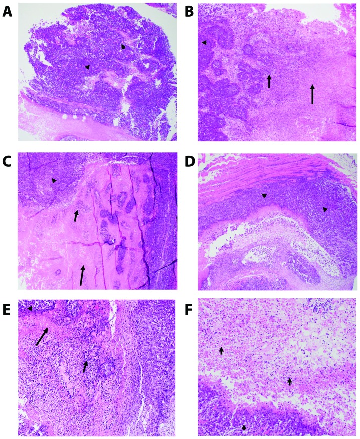
Figure 5.
(A-F) Photomicrographs of the mouse-bearing Ehrlich ascites carcinoma (EAC) tumor tissues. (A) Control untreated. Sheets of pleomorphic tumor cells (arrow head) (×4). (B) Yeast treated. Tumor (arrow head)/colliquative (middle arrow)/coagulative (right arrow) necrosis (×10). (C) Paclitaxel-H. Tumor (arrow head)/colliquative (middle arrow)/coagulative (tall arrow) necrosis (×10). (D) Paclitaxel-L. Sheets of pleomorphic tumor cells (arrow head) (×4). (E) Yeast + Paclitaxel-H. Tumor (arrow head)/colliquative (middle arrow)/coagulative (tall arrow) necrosis (×10). (F) Yeast + Paclitaxel-L. Tumor (arrow head), Colliquative necrosis (×10).

