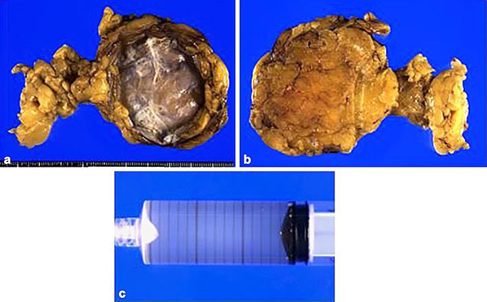
Fig. 3.

Macroscopic findings of the resected specimen. a The resected specimen was monolocular and mostly a fibrous cystic lesion with calcification of the cystic wall but without mural nodules. b Dorsal view of the specimen. c The content inside the lesion was a slightly mucinous yellowish fluid.
