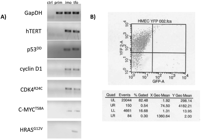
Fig. 2.
Confirming expression of transgenes. A) All transfections were validated by transgene confirmation using quantitative RT-PCR for the prim, imo, and tfo cell lines, and a “no template” control (ctrl). The imo and tfo cells were maintained in antibiotic-free media for 20–25 passages before reconfirming transgene expression. The final PCR products are shown. B) Transformed cells were sorted using FACS at p46. As seen in the table, the selected cells maintained over 80% fluorescence.

