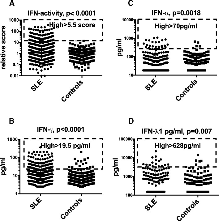
Fig. 1.
Type I IFN activity and levels of IFN-α, IFN-γ and IFN-λ1 in SLE patients and population controls. Type I IFN activity in vitro (a) and IFN-γ levels (b), IFN-α (c) and IFN-λ1 (d) were all higher in SLE patients then population controls (Mann-Whitney U test). The dashed boxes indicate individuals with high levels (> 75th percentile of patient measures) of each investigated IFN

