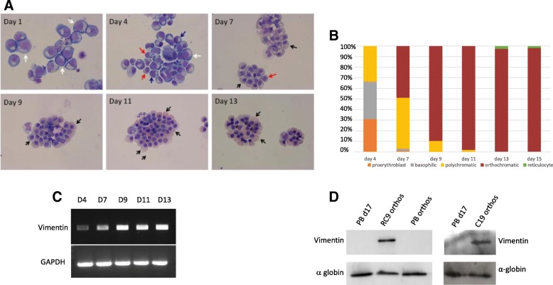
Fig. 2.
Expression of vimentin in erythroid cells differentiated from the ESC line RC9 RC9 CD34+ cells were incubated for up to 19 days in a three-stage erythroid culture system. a Cells were stained with May-Grünwald-Giemsa reagent at time points throughout the culture. White arrows, pro-erythroblasts; blue arrows, basophillic erythroblasts; red arrows, polychromatic erythroblasts; black arrows, orthochromatic erythroblasts. (b) The proportion of cells (Y-axis) at different stages of differentiation counted (data is representative of three cultures). c The abundance of vimentin transcripts at time points throughout erythroid culture was analysed by PCR. Abundance of GAPDH transcripts was used as a control. d Cells at day 17 in adult culture (orthochromatic erythroblasts and reticulocytes), isolated adult orthochromatic erythroblasts, RC9 and C19 orthochromatic erythroblasts were probed with an antibody to vimentin. Blots were stripped, and an α-globin antibody used as a control for protein loading

