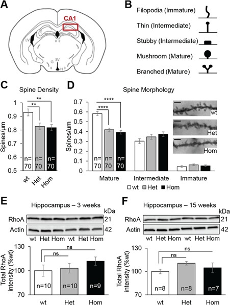
Figure 3.

Kctd13 deficiency induce spine maturation deficit in the CA1 region of the hippocampus. (A) Coronal brain diagram indicating the region of interest. (B) Categorization of Golgi-Cox-stained dendritic spine types. Ten micrometer stretches of dendrites were analyzed. (C) Quantification of dendritic spine density of CA1 pyramidal neurons. (D) Representative dendrite stretches (scale bar: 2 μm) and spine morphology of dendritic stretches analyzed. (E and F) Western blots against RhoA and β-actin. Hippocampi from 3- and 15-week-old mice were used as samples. Data are represented as the mean ± s.e.m.; ns, not significant; ****P < 0.0001 versus wt. Tukey’s test applied following a significant one-way ANOVA.
