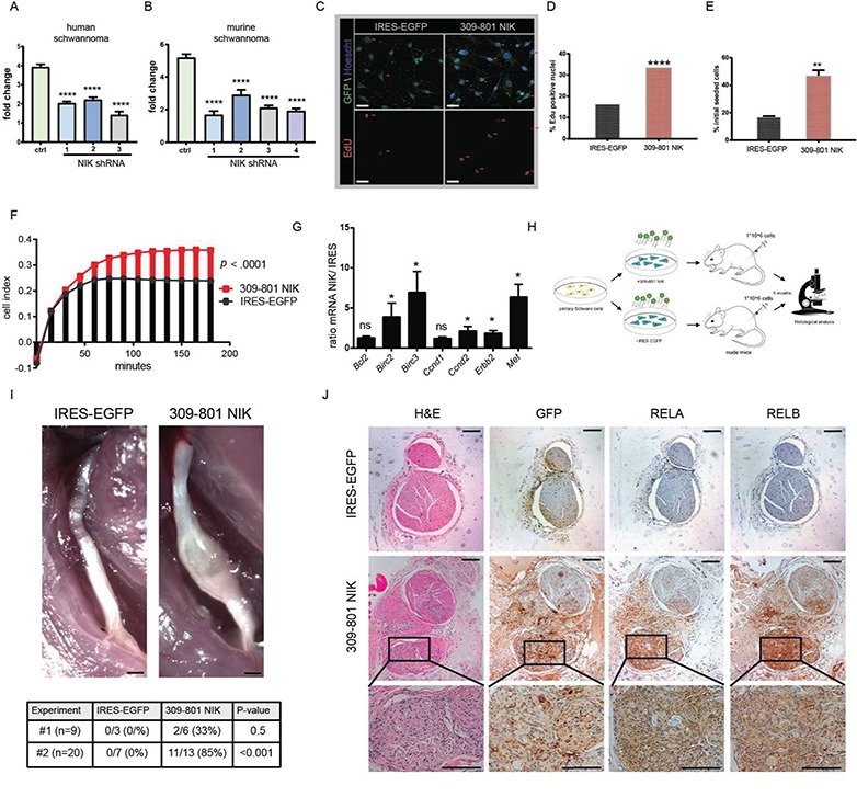
Figure 3.

NIK critically regulates SC function. (A) 72 h proliferation study in HEI-193 human schwannoma cell line comparing control and three independent NIK shRNAs. (B) 72 h proliferation study with primary murine schwannoma cells comparing control and four independent NIK shRNAs. In (C)–(J) primary passages 1 and 2 SCs were stably transduced with an IRES-EGFP control or 309-801 NIK IRES-EGFP expressing lentivirus. (C) Representative images of EdU immunofluorescence experiment. Scale bar = 20 μm. Original magnification ×200. (D) Quantitation of (C). Over 400 cells were counted of each genotype in total (n = 3 of each). (E) 48 h serum and growth factor starvation survival assay. (F) iCelligence cell adhesion assay. Adhesion is measured by changes in impedance caused by cells falling out of suspension and depositing onto electrodes, forming focal adhesion complexes. (G) qRT-PCR in transduced primary SCs. (H) Transplant schema of transduced primary SCs into Foxn1nu nude mice. (I, upper) Gross images of dissected sciatic nerves from IRES- and NIK-transduced SC transplants. Scale bar = 500 μm. (I, lower) Tabular results from transplant studies outlined in (H). (J) Serial sections of IRES- and NIK-injected nerves stained with H&E, GFP, RELA and RELB. Original magnification ×100, magnified inset 400×. Scale bar = 100 μm. [ANOVA with Dunnett’s test (α = 0.05) for post hoc comparisons (A) and (B); Fisher’s exact test (D); unpaired Student’s t-test with Holm–Sidak correction for multiple comparisons (α = 0.05, E, F, G, and I). ns = not significant, *P < 0.05, **P < 0.01, ****P < 0.0001. Error bars represent SEM. All experiments with statistical testing were performed in triplicate.]
