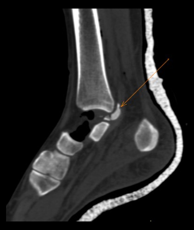
Figure 3.

Sagittal computed tomography image. Examination reveals extrusion of most of the right talus bone, with visualization of only the posterior fracture fragments which are dislocated posterosuperiorly (orange arrow) abutting the posterior malleolus. Emphysematous changes noted at ankle joint and surrounding soft tissues, with gas bubbles extending superiorly through the intramuscular fractures and subcutaneous tissues correlating with the presence of soft tissue laceration. Posterior cast is present.
