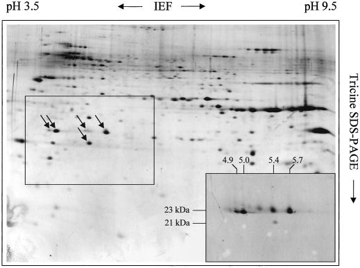
Figure 6.
Separation of multiple TOM20 forms from Arabidopsis. Total outer mitochondrial protein from Arabidopsis was separated by two-dimensional IEF/Tricine SDS-PAGE as described in “Materials and Methods.” The gel was silver stained. Some spots in the central frame specifically reacted with an antibody directed against TOM20 from potato (western blot in the right corner at the botton of the gel). The numbers on top and on the left of the western blot refer to the pI and molecular masses of the immunopositive spots.

