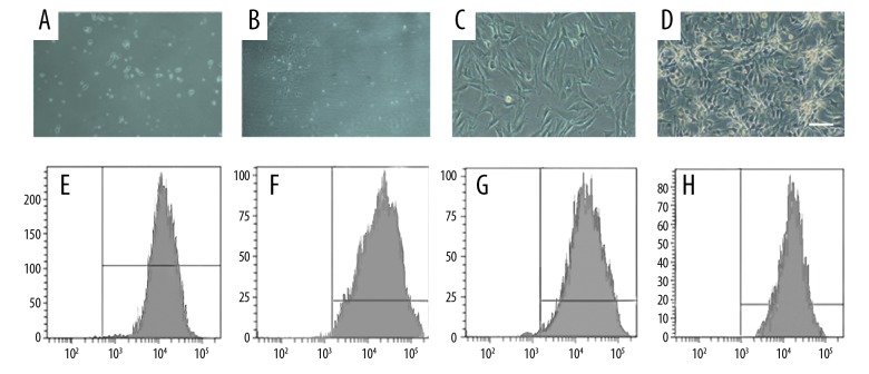
Figure 1.
Culture of BMSCs and identification of their surface molecular markers. (A) Spindle-shaped morphology of the marrow cells at day 1 (×10). (B, C) The number and size of the colonies gradually increased on days 3–5 (C×20, D×20). (D) A homogeneous population of BMSCs in the first passage of cultured marrow cells (×10). (E–H) Assay of cell surface antigens on rat mesenchymal stem cells.

