Figure 1.

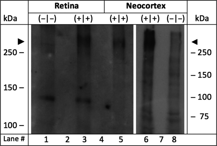
Expression analysis of Cav2.3 after SDS‐polyacrylamide gel electrophoresis and Western blot. Microsomal membranes were isolated from the retina and the neocortex of Cav2.3 null mutants and wild‐type littermates.12 Lane 1: 49 μg protein from Cav2.3‐deficient retina (−|−). Lane 3: 34 μg protein from Cav2.3‐competent retinae (+|+). Microsomal membranes from murine neocortices of Cav2.3‐deficient mice were used as negative (−|−) and from Cav2.3‐competent mice as positive controls (+|+), respectively. Lane 5: 32 μg protein from Cav2.3‐competent neocortical membranes. Lane 6 and 8, 72 μg of neocortical microsomal protein from Cav2.3‐competent or Cav2.3‐deficient mice, respectively. In lanes 2, 4, and 7, no protein was loaded. Peptide‐directed antibodies directed against a common epitope in all Cav2.3 splice variants (anti‐a1E‐com) were used as the primary antibody during Western blot analysis.9 Note that the predicted size of the full length Cav2.3 protein (262 kDa) is marked by the arrowheads
