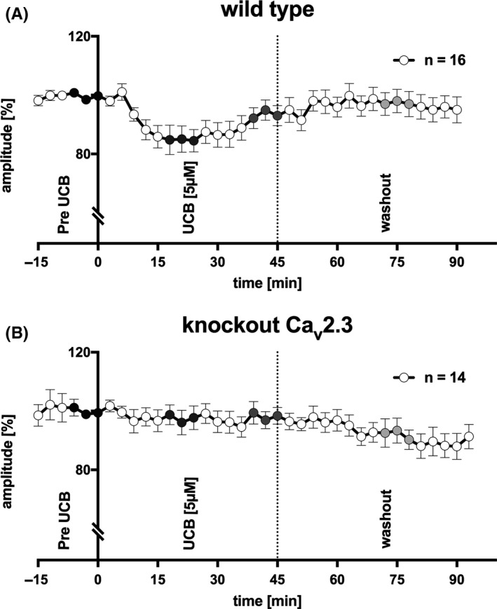
Figure 3.

ERG recordings from superfused murine retinae. (A) B‐wave amplitude in response to repetitive light stimulation every 3 min in wild‐type mice (n = 16). UCB was only added after reaching an equilibrium of b‐wave amplitude with albumin (pre‐UCB, black circles). Superfusion with UCB 5 μmol/L lasted 45 min and was followed by a washout of the retina by albumin only. UCB significantly decreased the b‐wave amplitude after approximately 18‐24 min (P < 0.05, corrected with Holm‐Bonferroni, dark gray circles), with partial recovery toward the end of UCB superfusion (P = 0.09, corrected, gray circles). After washout, the b‐wave amplitude recovered completely (n = 16; wild type, light gray circles). (B) UCB effects were not observed in Cav2.3‐deficient mice (n = 14). After washout, the b‐wave amplitude tended to decrease (P = 0.09, corrected)
