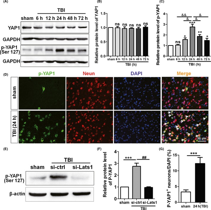
Figure 5.

YAP1 phosphorylation level is increased after TBI, and inhibition of Lats1 represses YAP1 phosphorylation. (A) Western blot detected the expression levels of YAP1 and p‐YAP1 at 6 h, 12 h, 24 h, 48 h, and 72 h following TBI. (B, C) The protein levels of YAP1 and p‐YAP1 were quantified as shown in (A), and the protein level of p‐YAP1 reached the peak at 24 hrs after TBI. (D) Immunofluorescence results indicated that p‐YAP1 (green) was expressed in rat cortical neurons (red). And the expression levels of p‐YAP1 were increased in TBI group. (E) Western blot analysis protein levels of p‐YAP1 in sham group, control siRNA group, and Lats1 siRNA group under the premise of silenced Lats1. (F) Quantification of the p‐YAP1 protein levels in each group as shown in (E). (G) Density of p‐YAP1 + cells. Scale bars: 50 μm. Values represent means ± SEM. Student's t test and one‐way ANOVA. *P < 0.05; **P < 0.01; ***P < 0.001 vs sham group; & P < 0.05; && P < 0.01 vs TBI group (24 h); ## P < 0.01 vs si‐ctrl group; ns: not significant
