
Figure 1.
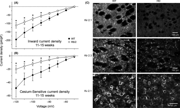
Reduction of inward K+ currents and Kir 2.1, 2.3 and Kv2.1 immuofluorescence intensity in R6/2 transgenic mice. (A‐B). Current‐voltage (I/V) relationships for inward K+ current densities (A) and Cs+‐sensitive K+ current densities (B) obtained from acutely dissociated MSNs from 11‐ to 15‐wk R6/2 transgenics and WTs. In these experiments, a 20 mM K+ concentration in the external solution was used to set the equilibrium potential at approximately −50 mV, corresponding to the holding potential. Obvious reduction of inward K+ current and Cs+‐sensitive K+ current occurred in R6/2 MSNs. (C). Immunofluorescent staining for the K+ channel proteins Kir2.1, Kir2.3 and Kv2.1. Top: Kir2.1 was detected within MSNs and the neuropil but was excluded from the fiber bundles of the internal capsule. The cellular fluorescent signal in R6/2 mice was decreased by 48%. Middle: Kir2.3 was visible within MSNs and the striatal neuropil but not found in fiber bundles. The cellular luminosity was decreased 26% in R6/2 compared with the WT mice. Bottom: Kv2.1 was associated with the membrane rim and initial processes of MSNs. Cellular luminosity decreased 47% in the HD compared with the WT. [Correction added on 19 February 2018, after first online publication: The word “Kv3.1” was changed to “Kv2.1” in the sentence “Reduction of inward K+ currents and Kir 2.1, 2.3 and Kv3.1 immuofluorescence intensity in R6/2 transgenic mice”.]