Figure 2.

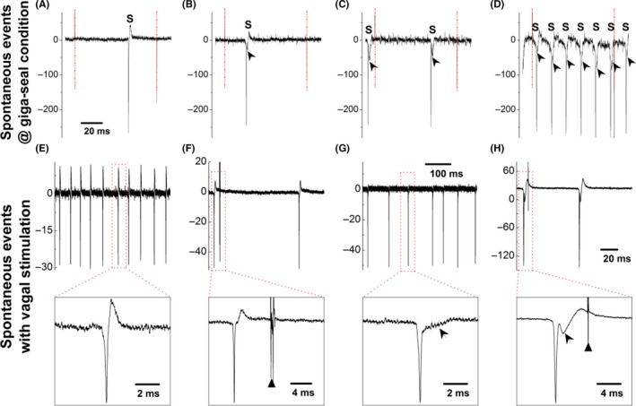
Spontaneous events (S) observed in myelinated A‐ (A) and Ah‐type (B‐D) baroreceptor neurons (BRNs) of nodose slice preparation using adult female rats under the giga‐seal condition with voltage‐clamp protocol before rupturing the cell membrane. Arrow heads represent the repolarization hump. The horizontal scale bar in (A) also applied for (A‐C). Dash‐dot line represents the seal pulse start and the end. Spontaneous events (S) recorded on myelinated A‐ (E) and Ah‐type (G) baroreceptor neurons (BRNs) under the current‐clamp mode without vagal stimulation and holding potential before rupturing the cell membrane; Spontaneous events (S) recorded on myelinated A‐ (F) and Ah‐type (H) BRNs under the current‐clamp mode with vagal stimulation (stimulus intensity below threshold) and without holding potential before the rupturing the cell membrane. Insets present the expended timescale to clearly demonstrate the repolarization hump (indicated as the arrowheads), black triangles present the time of vagal stimulation
