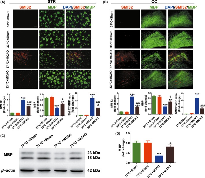
Figure 2.

Hypothermia promotes WM integrity and increases MBP protein levels at day 28 after MCAO. A and B, Representative images and quantitative data of MBP (green) and SMI32 (red) immunostaining in the striatum (STR, A) and corpus callosum (CC, B) in the ipsilateral hemisphere. Scale bar: 50 μm. Degree of WM injury was expressed as the fold increase in SMI32 (A & B, lower, left), decrease in MBP (A & B, lower, middle), and the relative ratio of SMI‐32 to MBP (A & B, lower, right) immunostaining intensity. C, Representative images of western blot performed using the ipsilateral brain tissues at day 28 in MCAO and sham groups. β‐actin was used as an internal control. D, Quantitative data showed that the MBP was significantly higher in hypothermia‐treated mice (33°C) than in normothermia mice (37°C); n = 5 per group. Data were expressed as mean ± SEM. *P < 0.05, **P < 0.01, ***P < 0.001 versus sham groups, #P < 0.05, ###P < 0.001 versus 37°C + MCAO group
