Figure 4.

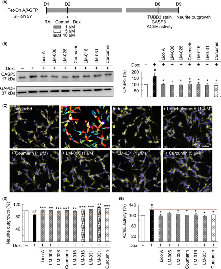
Neuroprotective effects of test compounds on Tet‐On Aβ‐GFP SH‐SY5Y cells. A, Experimental outline. Cells were plated with retinoic acid (RA, 10 µmol/L). On the following day, 1 µmol/L Lico A, coumarin, LM‐006, LM‐026, 1‐10 µmol/L LM‐016, LM‐031, or 5 µmol/L curcumin were added to cells for 8 hours, followed by the induction of Aβ‐GFP expression (+ Dox, 2 µg/mL) for 6 days. Neurite outgrowth was assessed after TUBB3 staining. In addition, the CASP3 level and AChE activity were assessed. B, CASP3 protein level was analyzed through immunoblotting using CASP3 and GAPDH (internal control) antibodies (n = 3). To normalize, the expression level in uninduced (−Dox) cells was set at 100%. P values between induced and uninduced cells as well as between treated and untreated cells were compared. C, Fluorescence microscopy images of uninduced or induced Aβ‐GFP cells treated with or without 1 µmol/L Lico A, coumarin, LM‐016, LM‐031, or 5 µmol/L curcumin. Shown next to the uninduced image is the image segmentation of uninduced cells with a multicolored mask to assign each outgrowth to a cell body for quantification. D, Quantification of neurite outgrowth with 1 µmol/L Lico A, coumarin, LM‐006, LM‐026, 1‐10 µmol/L LM‐016, LM‐031, or 5 µmol/L curcumin treatment (n = 3). To normalize, P values between induced and uninduced cells as well as between treated and untreated cells were compared, with the relative neurite outgrowth of uninduced cells as 100%. E, AChE activity assay with 1 µmol/L Lico A, coumarin, LM‐006, LM‐026, LM‐016, LM‐031, or 5 µmol/L curcumin treatment (n = 3). The relative AChE activity of uninduced cells was normalized to 100%. P values between induced and uninduced cells (# P < 0.05 and ## P < 0.01) as well as between treated and untreated cells (*P < 0.05, **P < 0.01, and ***P < 0.001) were compared
