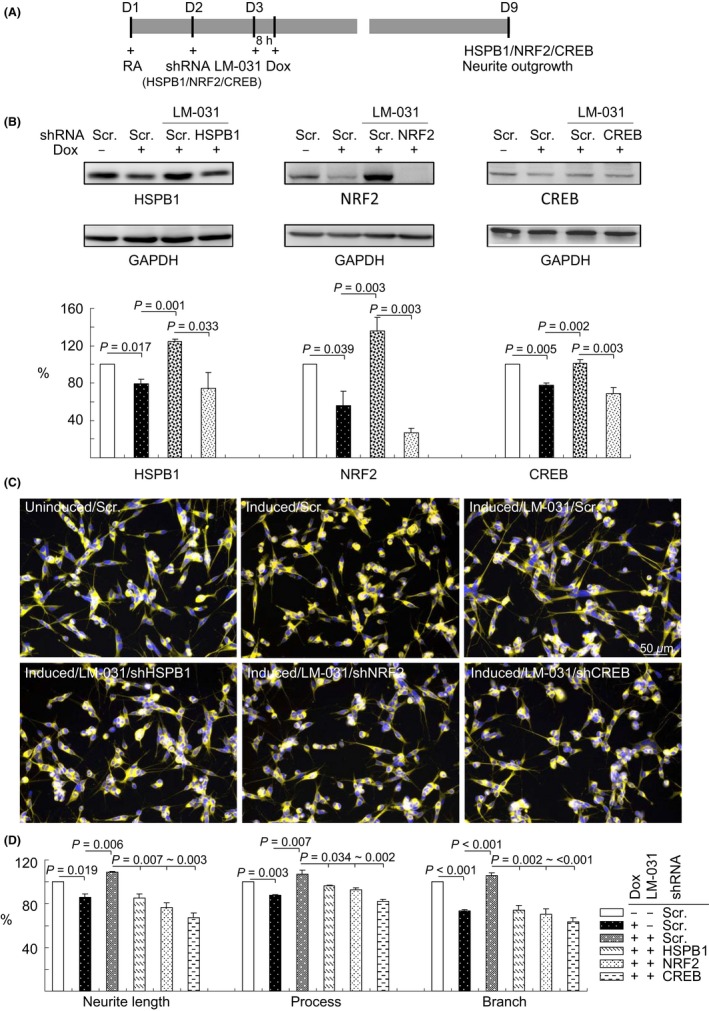
Figure 6.

HSPB1, NRF2, and CREB as therapeutic targets in LM‐031‐treated Aβ‐GFP SH‐SY5Y cells. A, Experimental outline. Aβ‐GFP SH‐SY5Y cells were plated into 6‐well (for protein analysis) or 24‐well (for outgrowth analysis) plates with retinoic acid (RA, 10 µmol/L) added on day 1. On the following day, cells were infected with lentiviruses expressing shRNA (HSPB1, NRF2, CREB‐specific, or scrambled). At 24 hours postinfection, LM‐031 (1 µmol/L) was added to the cells for 8 hours, followed by the induction of Aβ‐GFP expression (+ Dox, 2 µg/mL) for 6 days. Then, the cells were collected for HSPB1, NRF2, or CREB protein analysis through immunoblotting (GAPDH as a loading control) and neurite outgrowth analysis through high‐content analysis. B, Western blot analysis of HSPB1, NRF2, and CREB protein levels in LM‐031‐treated cells infected with HSPB1, NRF2, CREB‐specific, or a negative control scramble shRNA. To normalize, the relative HSPB1/NRF2/CREB level of uninduced cells was set at 100%. P values: comparisons between induced vs uninduced cells, LM‐031‐treated vs untreated cells, or scramble vs HSPB1/NRF2/CREB shRNA‐infected cells (n = 3). C, Microscopic images of uninduced or induced Aβ‐GFP SH‐SY5Y cells infected with scramble shRNA or LM‐031 (1 μmol/L) treated cells infected with scramble or HSPB1/NRF2/CREB‐specific shRNA. Nuclei were counterstained with Hoechst 33342 (blue). D, Neurite outgrowth assay, including length, process, and branch, of LM‐031‐treated Aβ‐GFP SH‐SY5Y cells infected with HSPB1/NRF2/CREB‐specific or a scramble shRNA. To normalize, the relative neurite length/process/branch of scramble shRNA‐infected, uninduced cells without LM‐031 treatment was set at 100%. P values: comparisons between induced vs uninduced cells, LM‐031‐treated vs untreated cells, or scramble shRNA vs HSPB1/NRF2/CREB‐specific shRNA‐infected cells (n = 3)
