Figure 4.

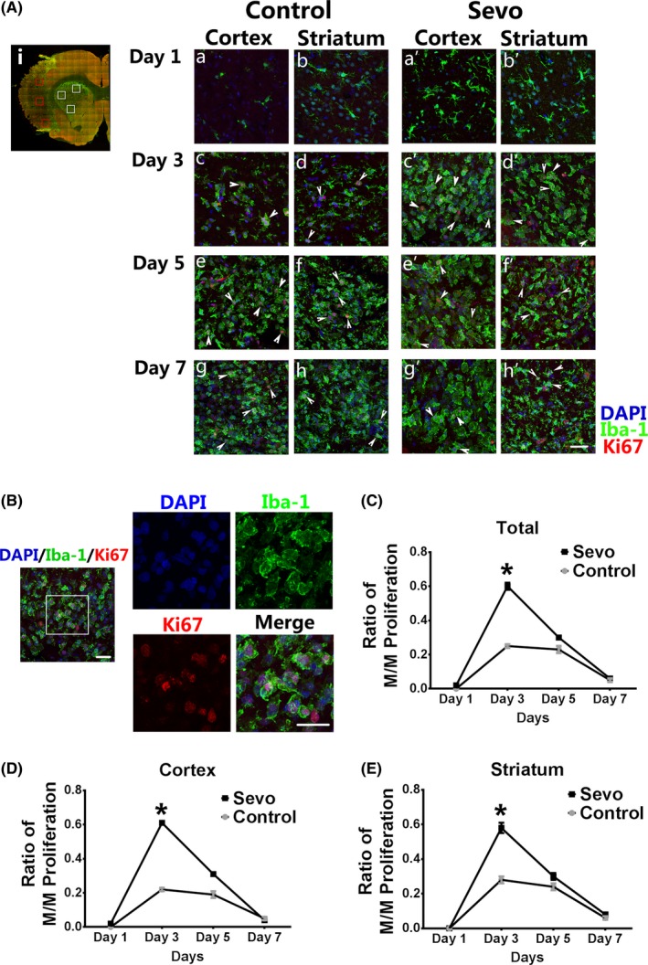
Sevoflurane preconditioning promoted microglia/macrophage proliferation after I/R injury. Ai: Representative stitching image shows the boxed areas for observation and calculation of proliferating microglia/macrophages (M/M) after injury. Aa‐h', Comparison of proliferating M/M stained with Iba1 and Ki67 in the infarcted regions between 2 groups from day 1 to day 7 after ischemia. Scale bar = 25 μm. White arrows show the proliferating M/M with Iba1 and Ki67 costaining. B, Representative confocal image shows the proliferating M/M. The boxed area indicates magnified region of the right panels. Scale bar = 20 μm. C‐E, The ratio of M/M proliferation was analyzed in the infarcted cortex and striatum from day 1 to day 7 after injury. Data were shown as mean ± SEM. *P < .05, n = 8
