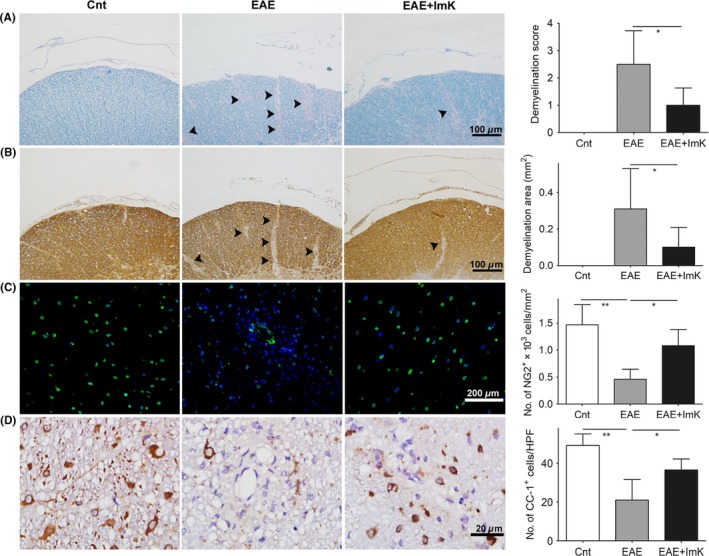
Figure 2.

The prevention of ImK on demyelination and oligodendrocyte loss in the Experimental autoimmune encephalomyelitis group. Luxol fast blue (A), immature oligodendrocyte (NG2) (B), and mature oligodendrocyte marker (CC‐1, MBP) (C, D) were stained in each group. (A, B): Arrows point to the border of the demyelinating area in the white matter. Quantification of demyelination scores (A, n = 6, *P < 0.05), demyelination area (μm2) (B, n = 8, *P < 0.05), and density of NG2+ OPCs (C, n = 6, *P < 0.05, **P < 0.01) and CC‐1+ oligodendrocytes (D, n = 6, *P < 0.05) were analyzed in the lumbar spinal cord white matter. HPF: 1000× high‐power field
