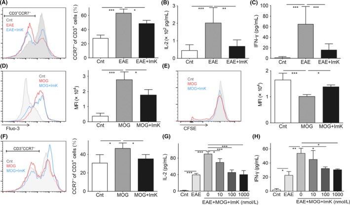
Figure 5.

Effects of ImK on peripheral T‐cell activation and cytokine release. CD3+ CCR7− cells (A) and serum IL‐2 (B) or IFN‐γ (C) from peripheral blood of each group after ImK treatment. The intracellular calcium concentration (D), proliferation (E), and CCR7− proportion (F) of T cells were quantified from restimulated PBMCs of Experimental autoimmune encephalomyelitis (EAE) rats by with or without ImK pretreatment. IL‐2 (G) or IFN‐γ (H) in the supernatant of control, EAE, or EAE PBMC restimulated by MOG 35‐55 with different concentrations of ImK was detected using ELISA. (Each group n = 3, *P < 0.05,**P < 0.01, ***P < 0.001)
