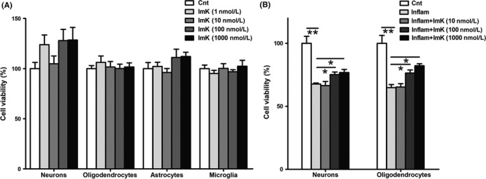
Figure 6.

Neural cell viability after exposure to ImK and ImK‐treated inflammatory supernatant. (A) Primary neurons, oligodendrocytes, astrocytes, and microglia viabilities were determined following treatment with varying concentrations of ImK ranging from 1 nM to 1 μM for 48 h (n = 4 for neurons, n = 6 for oligodendrocytes, n = 3 for astrocytes, n = 4 for microglia, P > 0.05 compared with the control). Neuron and oligodendrocyte viability (B) were quantified with exposure to different concentrations of ImK‐treated inflammatory supernatant for 48 h (n = 3, *P < 0.05,**P < 0.01)
