
Figure 4.
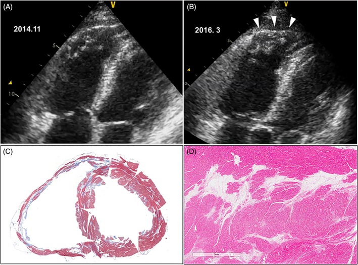
A and B, Difference in echocardiographic features 16 months apart. In addition to the slightly enlarged RV, the RV apex showed aneurysmal bulging during systole (arrowheads) mimicking ARVD. C, Explanted heart after cardiac transplantation. The myocardium stained blue (Masson trichrome stain) suggesting focal fibrosis. In addition, large areas of fatty tissue replacement mimicking ARVD are seen in the RV and left ventricle. D, High‐power view of fatty tissue replacement. ARVD, arrhythmogenic right ventricular dysplasia; RV, right ventricle