
Figure 5.
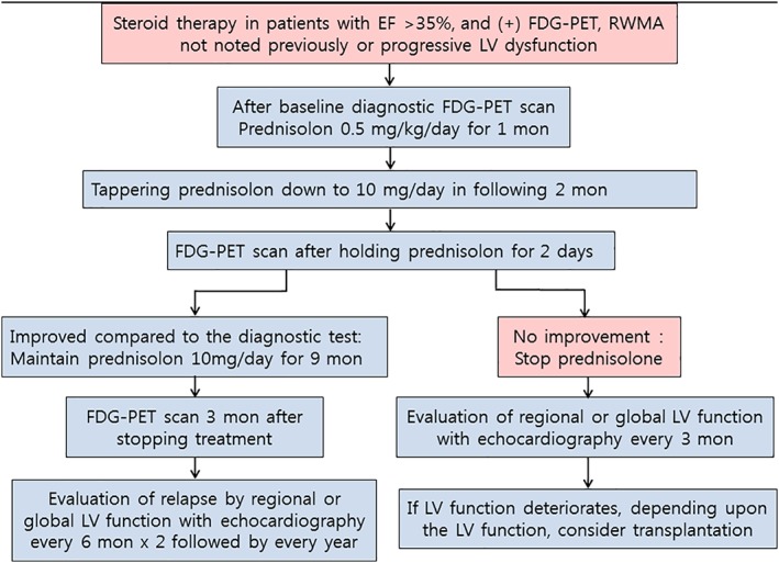
Treatment and follow‐up scheme. EF, ejection fraction; FDG‐PET, 18‐Fluoro‐2‐deoxyglucose positron emission tomography; LV, left ventricular; RWMA, regional wall motion abnormality
Official websites use .gov
A
.gov website belongs to an official
government organization in the United States.
Secure .gov websites use HTTPS
A lock (
) or https:// means you've safely
connected to the .gov website. Share sensitive
information only on official, secure websites.
Treatment and follow‐up scheme. EF, ejection fraction; FDG‐PET, 18‐Fluoro‐2‐deoxyglucose positron emission tomography; LV, left ventricular; RWMA, regional wall motion abnormality