
Figure 2.
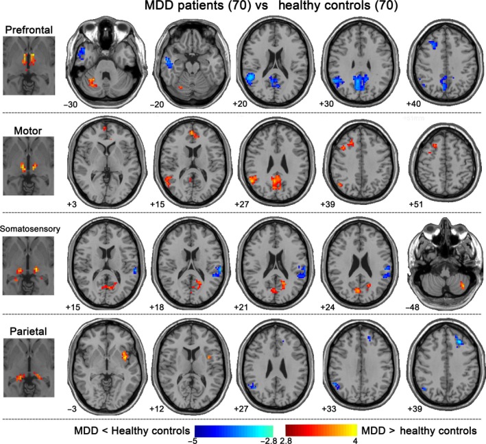
Thalamo‐cortical functional connectivity changes in MDD. Patients demonstrated reduced connectivity between prefrontal/parietal thalamus ROIs and bilateral MFG and right posterior DMN and between prefrontal/motor thalamus ROIs and lateral temporal regions. Conversely, increased connectivity emerged between motor thalamus ROI and right MFG and right medial frontal gyrus/anterior cingulate; between motor/somatosensory thalamus ROIs and right posterior DMN; between prefrontal/somatosensory thalamus ROIs and cerebellum; and between parietal thalamus ROI and left insula. All statistical maps were corrected for multiple comparisons using Gaussian random field theory with a cluster threshold of P < 0.05 and z > 2.9. MFG, middle frontal gyrus; DMN, default mode network; MDD, Major depressive disorder