
Figure 2.
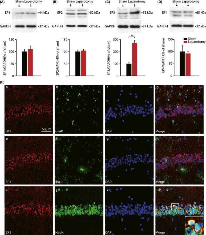
Protein levels of EP1‐4 in the hippocampus of postsurgery mice and cellular localization of EP3 expression. (A‐D) Western blot analysis demonstrated that laparotomy exclusively elevated hippocampal EP3 receptors expression at 7 days postsurgery, but not EP1, EP2, and EP3. (n = 3) (E) Immunofluorescence double staining showed that anti‐EP3 receptor staining colocalized with the neuron marker NeuN (i‐l), but not astrocyte marker GFAP (a‐d), nor microglia marker Iba‐1 (e‐h) in the CA1 of the hippocampus 7 days after laparotomy. Nuclei were stained with DAPI. (Scale bars: 50 μm). Data presented as mean ± SEM. **P < 0.01 [Correction added on 03 April 2018, after first online publication: The word “GADPH” was changed to “GAPDH” in figure 2.]