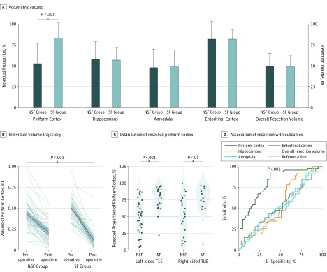
Figure 2. Volumetric Results in the Derivation Cohort of Patients Undergoing Surgery for Temporal Lobe Epilepsy (TLE).
Volumetric results (A) are displayed as median (SD) (error bars) of the resected proportion of the piriform cortex, hippocampus, amygdala, and entorhinal cortex and the overall resection volume in the overall cohort (n = 107). Comparing postoperatively seizure-free (SF [n = 46]) with non–seizure-free (NSF [n = 61]) groups, difference in piriform cortex volumes was significant, whereas no differences were observed in all other regions. Gray lines show the individual trajectories of presurgical vs postsurgical volumes of the piriform cortex in the SF vs NSF groups (B). The mean trajectory is illustrated by a dark blue line; SD, by the light blue area. The distribution of the resected proportion of the piriform cortex in the NSF vs SF groups is shown (C); the 50% resection cutoff is displayed as a blue horizontal line. The association of resection area with outcomes (D) used receiver operating characteristics curves to describe individual discrimination. The piriform cortex was the only area to significantly discriminate between SF and NSF groups in the derivation cohort. All other areas remained close to the 45° reference line.

