
Figure 2.
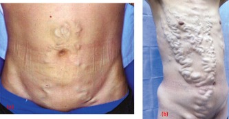
(A) A caput Medusæ of mildly tortuous abdominal wall veins containing hepatofugal blood and radiating from the umbilicus, in a patient with cirrhosis and portal hypertension. The caput Medusæ results from backflow from the left portal vein through paraumbilical veins in the falciform ligament, to periumbilical systemic veins in the abdominal wall. Reproduced with permission from New England Journal of Medicine.24 Copyright 2005, Massachusetts Medical Society. (B) Abdominal wall varices caused by thrombosis of the suprahepatic and posthepatic portions of the inferior vena cava, in a patient with thrombosis of the hepatic veins (Budd‐Chiari syndrome). Reproduced with permission from New England Journal of Medicine.25 Copyright 2014, Massachusetts Medical Society.