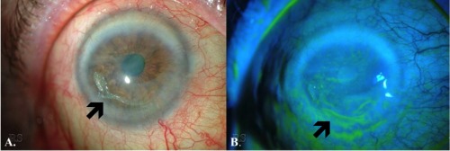
Figure 3.

Slit‐lamp photos of a patient with severe, complex surface disease secondary to keratoconjunctivitis sicca. (A) Conjunctival injection, diffuse haze throughout the cornea, corneal neovascularization, and epithelial erosions are evident. (B) Note the epithelial erosions and irregularity shown with fluorescein staining.
