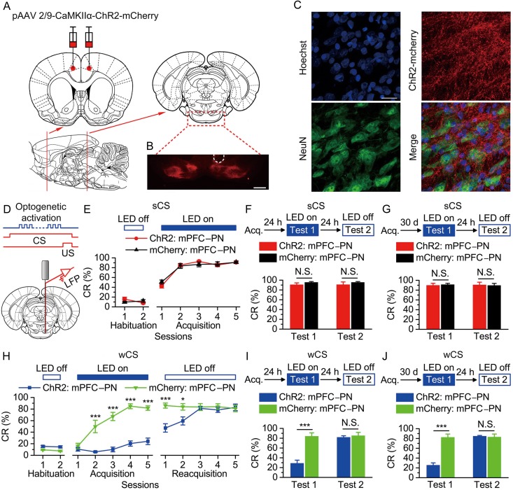
Figure 5.
Optogenetic activation of the bilateral caudal mPFC axon terminals in the right PN abolished the acquisition, recent and remote retrieval of DEC with the wCS, but not of DEC with the sCS. (A) Rats were bilaterally injected with pAAV 2/9-CaMKIIα-ChR2-mCherry or pAAV 2/9-CaMKIIα-mCherry into the caudal mPFC. (B) Example of robust ChR2-mCherry expression in the axon terminals of mPFC in the PN. White dashed line: optrode position. Scale bar, 500 μm. (C) Representative images showing images of ChR2-mCherry -positive axons (red) innervating target neurons in the PN (NeuN, green). Scale bar, 20 μm. (D) Coronal schematic of optical fiber implant site and 470-nm LED illumination pattern to the caudal mPFC axon terminals in the PN during each trial. (E–J) Top: Training and illumination phase protocol. Bottom: Effects of optogenetic activation of mPFC axon terminals in the PN during each trial on CR% of acquisition (E: n = 10 each; H: n = 12 ChR2: mPFC–PN, n = 11 mCherry: mPFC–PN), recent retrieval (F: n = 11 ChR2: mPFC–PN, n = 10 mCherry: mPFC–PN; I: n = 12 ChR2: mPFC–PN, n = 11 mCherry: mPFC–PN), and remote retrieval (G: n = 10 ChR2: mPFC–PN, n = 11 mCherry: mPFC–PN; J: n = 12 for ChR2: mPFC–PN, n = 10 mCherry: mPFC–PN) of DEC with the sCS or wCS (N.S., not significant, *P < 0.05, ***P < 0.001; 2-way ANOVA with repeated measures followed by Tukey post hoc test or 2-tailed unpaired Student's t-test). Data are represented as mean ± s.e.m.

