
Figure 3.
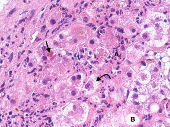
AH with coexisting AFD. This field shows the characteristic features of AH, with liver cell ballooning (curved arrow), Mallory‐Denk body formation (straight arrow), and neutrophilic infiltrates (A). Macrovesicular steatosis is present (B) but involves only a few hepatocytes. Microvesicular foamy hepatocytes were present in other fields of the biopsy.