
Figure 3.
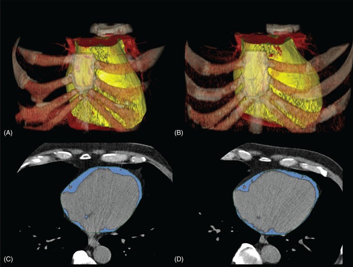
Volume rendered non–contrast‐enhanced native CT images of the chest of a 67‐year‐old male monozygotic twin pair (A, B). The yellow volume represents the epicardial adipose tissue compartment. The panels C and D are axial CT images of the same twin pair. The blue areas represent the epicardial adipose tissue compartments. Abbreviations: CT, computed tomography.