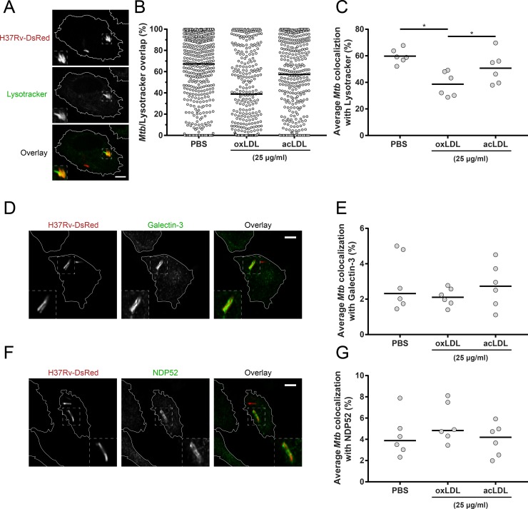
Fig 6. oxLDL impairs Mtb localization to lysosomes in macrophages.
Primary human macrophages were treated overnight with PBS control, acLDL or oxLDL (25 μg/ml) and subsequently infected with DsRed-Mtb H37Rv (red) at a MOI of 10:1. Cells were stained (green) for lysosomes (Lysotracker) (A), galectin-3 (D) or NDP52 (F) at 4 h post-infection and analyzed by confocal microscopy. Pictures were taken at a 63x magnification. Scale bars represent 5 μM. Percentage overlap of intracellular mycobacteria with staining was determined for 3 wells * 3 = 9 pictures per condition. (B) Results of a representative donor of Mtb overlap with Lysotracker. Individual mycobacteria are represented by dots with group medians. Average colocalization of Mtb with Lysotracker (C), galectin-3 (E) and NDP52 (G) are displayed for macrophages from six independent donors. Individual donors are depicted as dots with group medians. Statistical significance was determined by Wilcoxon signed rank test with post-hoc FDR correction. * = p < 0.05.

