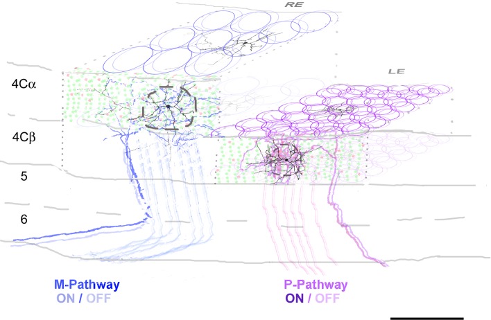
Figure 4.
Schematic representation of the organization of layer 4C in V1. Two ocular dominance columns (ODC’s) are shown as an example (RE and LE: right and left eye, respectively). For clarity only the M-pathway input is shown to the RE ODC and the P-pathway input to the LE ODC. We estimated that 20 and 203 TC fibers are distributed to each ODC within layers 4Cα and 4Cβ, respectively (solid blue and purple circles). We assumed an equal distribution of ON and OFF center afferents, shown as the slightly offset circle pairs. The fine-scale topographic arrangement of neighboring afferents within a column is not known, so the depiction illustrates only the sizes, not the arrangement. Within each ODC we calculated that there were 8 and 7 TC afferents within the dendritic field domain of each spiny stellate cell in 4Cα and 4Cβ, respectively. For each column the spatial extent of the axonal arbor of ON and OFF TC input is shown. The dashed circles indicate the approximate extent of the dendritic domains of spiny stellate neurons. Reconstructions of Golgi stained neurons and horseradish peroxidase-filled TC axons modified from Lund (1973) and from Blasdel and Lund (1983), respectively. Within layer 4Cα and 4Cβ there are 8185 and 14 315 neurons/ODC respectively; 87% are excitatory (green dots) and 13% inhibitory (red dots). Scale bar: 200 μm.

